International Journal of Pharmaceutical and Phytopharmacological Research
ISSN (Print): 2250-1029
ISSN (Online): 2249-6084

Moringa oleifera (M. oleifera) and Moringa concanensis (M. concanensis), belong to the family of Moringaceae and are well-known as the drumstick tree. M. oleifera is a miracle tree, that people have been using for centuries due to its health benefits. M. concanensis locally known as Kattumurungai is also one of the important medicinal plants. Like M. oleifera, this plant is also used to treat skin tumors, fatigue, high blood pressure, jaundice, and diabetes. The present study involved the comparative pharmacognostic and phytochemical evaluation of these two species. The preliminary phytochemical analysis of both species showed the presence of alkaloids, glycosides, flavonoids, steroids, and tannins including carbohydrates, proteins, and lipids. The maximum percentage yield was found in M. oleifera. The physicochemical parameters like ash values and extractive values were found to be maximum in M. oleifera. The thin layer chromatography (TLC) of the methanolic extract was performed for three important phytochemicals such as alkaloids, steroids, and flavonoids. Both species showed the presence of prominent spots on TLC plates. Among these spots, some of them had similar Rf values, but some of them with different Rf values indicating the presence of similar and different alkaloids, steroids, and flavonoids respectively. Determination of total flavonoid and total phenolic content was also carried out using the Folin Ciocalteu and Aluminum Chloride methods respectively. M. oleifera was found to contain 453 µg/ml of total phenols and 365 µg/ml of total flavonoids and were maximum compared to M. concanensis. These findings suggest that M. concanensis shares some similar macroscopical and phytochemical characteristics as that of M. oleifera but is very similar in the types of alkaloids, steroids, and flavonoids. Further study has to be done to identify these particular spots using reference standards.
INTRODUCTION
The Moringaceae family includes various species of Moringa among which Moringa oleifera (M. oleifera) and Moringa concanensis (M. concanensis) are two unique species. Each possesses unique botanical characteristics, phytochemical profiles, and traditional uses. The species have gained attention for their diverse array of bioactive compounds and potential health benefits, contributing to their recognition as valuable resources in various fields [1, 2].
M. oleifera is a well-known plant. It is often referred to as the "drumstick or miracle plant" and is native to Pakistan, Afghanistan, India, and Bangladesh [3, 4]. Renowned for its adaptability to different climates, Moringa oleifera is celebrated for its rich nutritional content, including vitamins, minerals, antioxidants, and essential amino acids. The plant's leaves, seeds, and other parts have been traditionally utilized in herbal medicine and culinary practices [5, 6].
On the other hand, M. concanensis is one of the lesser-known plants. It is a wild variety of moringa commonly known as the "West Indian Moringa", "Monga," or Kattumurugai and is native to the Western Ghats of India. While sharing the genus with M. oleifera, M. concanensis exhibits distinct botanical features and a unique chemical composition [7, 8].
Both species have been recognized for their potential medicinal properties and are being used in traditional medicine by local communities [9-11].
As we explore the comparative aspects of M. oleifera and M. concanensis it becomes evident that these plants offer a fascinating juxtaposition in terms of geographical distribution, morphological characteristics, and ethnobotanical significance. The species contains various phytoconstituents such as terpenoids, alkaloids, tannins, steroidal aglycones, and reducing sugars in different parts such as leaves, bark, flowers, seeds, roots, and pods [12, 13]. The species have been shown to exhibit anti-urolithic [14], antimicrobial [15], anti-bacterial [8, 16, 17], analgesic, anti-inflammatory [18-20], antioxidant [21-23], anti-diabetic [24, 25], hepatoprotective [26] and more activities. In the present study, we focused on comparing various pharmacognostic parameters including identification of phytochemicals, determination of physicochemical parameters, TLC of the extracts, and determination of total phenolic and flavonoid content of leaves of M. oleifera and M. concanensis.
MATERIALS AND METHODS
The plants, M. oleifera and M. concanensis were collected from the agricultural field near Miyapur, Hyderabad, and identified and authenticated by a botanist, Govt. Degree College, Kukatpally, Medchal district, Hyderabad.
Macroscopic study
The collected plant material of M. oleifera and MC were subjected to evaluation of morphological and organoleptic characters.
Preparation of the extract
The leaves were separated and were dried at room temperature under shade, by spreading on the filter paper and ground into coarse powder in a grinder. 250 gm powdered coarse material was extracted with methanol using a Soxhlet apparatus for 2 hrs. Then the extract was concentrated by evaporating in a water bath and the % yield was calculated [27]. The extract was subjected to various qualitative and quantitative analyses.
Preliminary phytochemical study
Detection of sugars
Molisch’s test
After treating the extract with an alcoholic α-Naphthol solution, conc. sulfuric acid was applied from the test tube's sides. The presence of sugars is indicated by the creation of a reddish-violet ring.
Fehling’s test
The extract was hydrolyzed with dilute hydrochloric acid and Fehling’s A and B solution were added. After heating the formation of a reddish brown colour indicates the presence of reducing sugars.
Detection of proteins
Biuret test
The extract was treated with a few ml of Biuret reagent. The pink or purple color obtained after heating the solution indicates the presence of proteins.
Ninhydrin test
To the extract few ml of Ninhydrin reagent was added and heated in a water bath. The presence of pinkish-red color shows the presence of proteins.
Detection of fixed oils and fats
Small quantities of extract were rubbed between the two filter papers and checked for oil stains [28, 29].
Detection of saponins
Froth test
0.1 g plant powder was vigorously shaken with a few ml of distilled water for five min and was allowed to stand and checked for froth after 15 minutes.
Detection of flavonoids
Shinoda test
A small amount of magnesium ribbon pieces and a small amount of concentrated hydrochloric acid were added to the extract. The development of the magenta hue denotes a favorable response to flavonoids.
Alkali test
The extract was treated with a dilute ammonia solution and the ammonia layer acquired a yellow colour showing the presence of the flavonoid.
Detection of steroids
Salkowski test
Few drops of conc. sulphuric acid was added to the chloroform extract and observed for the red color or golden yellow color in a lower layer which indicates the presence of the sterols or triterpenes respectively [30].
Liebermann–Burchard test
To the chloroform extract few drops of acetic anhydride were added followed by the addition of 1 ml of conc. sulphuric acid from the sides of the test tube [31].
Keller-Kilinai test
To the extract, 3 ml of acetic acid and a few drops of ferric chloride were added and this mixture was transferred to a test tube containing conc. sulphuric acid. A reddish brown ring turns bluish green indicating the presence of deoxy sugars.
Detection of alkaloids
Dragendroff’s reagent test
To the extract few drops of Dragendroff’s reagent were added and a reddish brown ppt indicates the positive test alkaloids.
Hager’s reagent test
To the extract, a few drops of Hager’s reagent were added and the formation of the yellow ppt is an indication of the presence of alkaloids [32].
Detection of tannins
Ferric chloride test
The methanolic extract was shaken with water and filtered. The filtrate was treated with an aqueous solution of ferric chloride appearance with a blue or brownish-green color indicating the presence of tannins.
Lead acetate test
The extract was shaken with water and treated with 10% lead acetate. The creamy yellow ppt or white ppt shows the presence of tannins [33-35].
Determination of physicochemical parameters
M. oleifera and MC samples were subjected to various physicochemical parameters as follows [36, 37].
Total ash value
About 2 g of the powder drug was added to the previously weighed silica crucible and ignited at the temperature of 500- 600 °C in a muffle furnace (Proto-tech, Mumbai) until white color ash was obtained. The crucible was reweighed and % of total ash content was calculated on a dried basis.
Acid-insoluble ash
To the total ash, 25 ml of dilute HCL was added and boiled gently. It was filtered through ashless filter paper and the residue left on the filter paper was washed with hot water until the filtrate showed neutral. Then the insoluble residue was burned to constant weight and reweighed. Acid-insoluble ash content was calculated.
Alcohol-soluble extractives
5 gm of the powdered drug was macerated with 100 ml of 90% alcohol and kept for 24 hrs with frequent shaking for 6 hrs. After 18 hr of standing, it was filtered and 25 ml of the filtrate was evaporated to dryness in preweighed porcelain, and the percentage content of alcohol-soluble extractive value was calculated based on the air-dried plant material.
Water soluble extractives
Chloroform water was used to determine water-soluble extractives following the same procedure mentioned under alcohol-soluble extractives instead of 90% alcohol.
Determination of petroleum ether soluble extractive values
The same procedure was followed as mentioned under alcohol-soluble extractive value. Petroleum ether was used instead of 90% alcohol.
Thin layer chromatographic study (TLC)
TLC was performed on the handmade Silica gel G plates. Using mobile phases, Toluene: Methanol (9:1) (Steroids), Toluene: Diethylamine: Ethyl acetate: (7:1:2) (Alkaloids), and Ethylacetate: Glacial acetic acid: Formic acid: Water (100:11:11:20) (Flavanoids) for the development of plates.
Preparation of sample
About 1 mg of extract of the M. oleifera and MC were dissolved in methanol for alkaloids, for flavonoids extract was dissolved in 80% methanol and for steroids, petroleum ether was used to dissolve the extract and applied on the TLC plates.
After the spotting of the sample, the plates were kept in the mobile phase and the spots were viewed in the UV chamber at 245 nm or 365 nm. The Rf values were calculated by dividing the distance traveled by the sample by the distance traveled by the solvent [38-40].
Determination of total phenol content
A standard calibration curve was made with Gallic acid. From the stock solution (100 μg/ml) of gallic acid, the different concentrations (10-50 μg/ml) were prepared 1 ml solution of each concentration was pipetted into a volumetric flask and diluted with 3 ml of distilled water. Then 0.5 ml of phenol reagent was added followed by the addition of 2 ml of Na2CO3 (2%) in each volumetric flask. The final volume was adjusted to 10 ml with distilled water. Absorbance was measured using a UV-visible spectrophotometer at 765 nm (Shimadzu UV-1800, Japan) against blank, containing distilled water.
For the sample preparation, 100 mg of M. oleifera and MC extracts were dissolved in 50 ml of 50% methanol in separate test tubes and filtered. One ml of each filtrate was pipetted in a 10 ml volumetric flask and proceeded with similar steps as mentioned in the preparation of the standard solution. Estimation of total phenols was done based on the calibration curve of gallic acid [41].
Determination of total flavonoid content
Total flavonoid content was determined by the Aluminum chloride method using quercetin to make a standard calibration curve. The different concentrations (10-50 μg/ml) were prepared from the stock solution (100 μg/ml) of quercetin. 1 ml from each of the solutions was pipetted out in a volumetric flask and 0.2 ml of 10% aluminum chloride, 3 ml of 95% methanol, and 0.2 ml of potassium acetate (1 M) were added and mixed well. Finally, the solutions were diluted to 10 ml with distilled water and kept at room temperature for 30 minutes. The absorbance of the resulting solutions was measured with a UV spectrophotometer at 415 nm.
For the sample preparation, 100 mg of M. oleifera and M. concanensis extracts were dissolved in 80% methanol and 1 ml of each was pipetted in 10 ml of a volumetric flask and proceeded with similar steps as mentioned in the preparation of the standard solution. Estimation of total flavonoid was done based on the calibration curve of quercetin [42].
RESULTS AND DISCUSSION
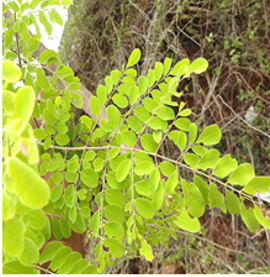
Morphological and organoleptic characters of M. oleifera (Figure 1a) and M. concanensis (Figure 1b) are given in Table 1.
Table 1. Morphological and organoleptic characters
|
Macroscopic characters |
M. oleifera |
M. concanensis |
|
Leaves |
Tripinneately compound |
Bipinneately compound |
|
Shape |
Oval |
Obovate |
|
Size |
1.4-5 cm |
2.2-5 cm |
|
Texture |
Smooth to touch |
Hard to touch |
|
Fruits |
Long, slender, and green |
Long and slender and green with brownish tinge |
|
Color |
Light green color |
Dark green color |
|
Odor |
Unpleasant odour |
Unpleasant odour |
|
Taste |
Bitter |
Bitter |
|
|
|
a) |
|
|
|
b) |
|
Figure 1. a) M. oleifera, and b) M. concanensis |
Preliminary phytochemical study
The medicinal and therapeutic activity of the plant is due to the presence of various phytochemicals and preliminary phytochemical study will help in the identification of these bioactive groups. In this study, the extract of M. oleifera and M. concanensis showed the presence of carbohydrates, lipids, proteins alkaloids, flavonoids, tannins, saponins, and steroids. However, the Kellar-Kiliani test was positive in M. oleifera and negative in the case of M. concanensis.
Physicochemical study
Physicochemical parameters are rarely constant but very important for the evaluation of medicinal plants as they indicate their identity, purity, and quality [43]. Ash values represent the inorganic content naturally present or adhering or deliberately added to it, as a form of adulteration. Extractive values are indicative of the approximate content of chemical constituents [28, 37]. The extract was subjected to various physicochemical parameters and results are given in Table 2. The Ash values and extractive values were found to be maximal in M. oleifera than in M. concanensis. The alcohol extractive values were found to be maximum followed by water and petroleum ether extractive values in both M. oleifera and M. concanensis.
Table 2. Physicochemical parameters
|
Physicochemical parameters |
w/w (%) ± SD (*n = 3) |
|
|
M. oleifera |
M. concanensis |
|
|
Total ash |
4.5 ± 0.42 |
3.5 ± 0.23 |
|
Acid insoluble ash |
1.1 ± 0.22 |
0.8 ± 0.35 |
|
Water soluble extractives |
18.7 ± 0.39 |
16.5 ± 0.43 |
|
Alcohol soluble extractives |
27.2 ± 0.13 |
25.5 ± 0.26 |
|
Petroleum ether-soluble extractives |
5.3 ± 0.21 |
4.9 ± 0.32 |
Note, *n = number of readings
Thin layer chromatographic study
TLC study is one of the simple methods in the identification of phytochemicals as It gives the fingerprint of constituents present in the drug sample. Based on the results one can have an idea about the isolation of phytoconstituents [44]. It has some benefits, including reducing analysis time and sample cost and being able to analyze numerous samples at once with a small amount of mobile phase [45, 46]. TLC coupled with various analytical detection systems can be effectively used for qualitative and quantitative separation of various crude mixtures [47].
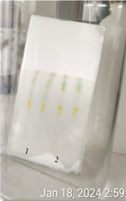
TLC analysis of M. oleifera and M. concanensis was carried out in three solvent systems Toluene: Methanol (9:1), Toluene: Diethylamine: Ethyl acetate: (7:1:2) and Ethylacetate: Glacial acetic acid: Formic acid: Water (100:11:11:20) which are specific for the separation of steroids, alkaloids and flavonoids and results are given in Table 3. For steroids, M. oleifera gave 4 spots with brownish to pink color fluorescence, and M. concanensis gave 6 spots with pinkish fluorescence under UV 365 nm (Figure 2), and in visible light, both species were found to show light brown to greenish color spots, but 3 spots were observed in M. oleifera and 4 in MC.
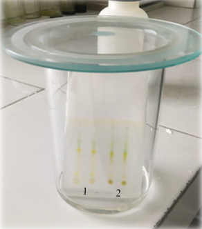
For alkaloids, both the extracts of M. oleifera and MC gave 4 spots under UV 365nm with pink color fluorescence (Figure 3) and in visible light, 5 spots of light brown to green color were observed in both the species having the same Rf values.
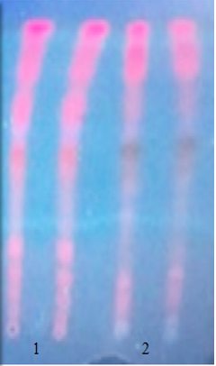
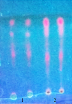
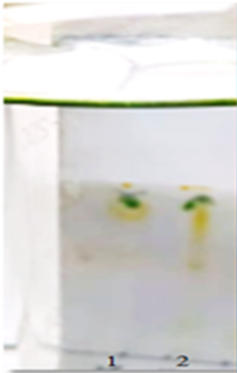
For flavonoids, M. oleifera gave 8 spots and M. concanensis gave 5 spots with blue, pink, and purplish fluorescence under UV 365 nm (Figure 4), and in visible light, 3 spots of light brown to greenish color were observed in both species.
Table 3. TLC study of M. oleifera and M. concanensis
|
Phytochemical groups |
Rf values |
|
|
M. concanensis |
M. oleifera |
|
|
Steroids (Figure 2) |
0.16, 0.28, 0.32, 0.44, 0.59, 0.76, 0.85 (7) |
0.24, 0.28, 0.40, 0.73, 0.83 (5) |
|
Alkaloids (Figure 3) |
0.54, 0.59, 0.70, 0.82 (4) |
0.54, 0.59, 069, 0.82 (4) |
|
Flavonoids (Figure 4) |
0.12, 0.55, 0.66, 0..80, 0.92 (5) |
0.17, 0.23, 0.27, 0.34, 0.45, 0.66, 0.80, 0.92 (8) |
|
|
|
|
a) |
b) |
|
Figure 2. TLC of steroids: a) In developing chamber, and b) Under UV 365 nm. Track 1: M. concanensis, track 2: M. oleifera |
|
|
|
|
|
a) |
b) |
|
Figure 3. TLC of alkaloids: a) In developing chamber, and b) Under UV 365 nm. Track 1: M. concanensis, and track 2: M. oleifera |
|
|
|
|
|
a) |
b) |
|
Figure 4. TLC of flavonoids: a) In developing chamber, and b) Under UV 365 nm. Track 1: M. concanensis, and track 2: M. oleifera |
|
Total phenols and total flavonoid content
The phenols and flavonoids are the most abundant phenolic compounds found in plants. İt is necessary to quantify the presence of these metabolites in the plants as they possess a wide array of pharmacological activities such as anti-inflammatory, antibacterial, anticancer antioxidant, etc. [41, 42]. Total Phenols and total flavonoids were determined using Folin Ciocalteu’s reagent method and the Aluminium chloride method in the extract, respectively.
The standard calibration plot of Gallic acid and Quercetin was constructed in the concentration range of 10-50 µg/ml. The coefficient of determination (R2) was found to be 0.991 and 0.998 for gallic acid and quercetin respectively. The extract of M. oleifera was found to contain maximum total phenols and flavonoids compared to M. concanensis. M. oleifera was found to contain 453 µg/ml of total phenols and 365 µg/ml of total flavonoids and M. concanensis was found to contain 393 µg/ml and 263 µg/ml of total phenols and total flavonoids respectively.
CONCLUSION
M. oleifera and M. concanensis, are the two important species of genus ‘Moringa’. M. oleifera has been studied extensively for its phytochemical content and pharmacological activities than M. concanensis. Thus the present was carried out to compare these two species. It is concluded that the the phytochemicals identified in both species were similar except the some. The phenols and flavonoids were found to be abundant in both species which are responsible for the antioxidant activity. Further study has to be done to isolate the active constituents of M. concanensis and evaluate their biological activities which will help treat the diseases.
Acknowledgments: We are thankful to the institution and management of Gokaraju Rangaraju College of Pharmacy, Hyderabad, for providing lab facilities.
Conflict of interest: None
Financial support: None
Ethics statement: None