International Journal of Pharmaceutical and Phytopharmacological Research
ISSN (Print): 2250-1029
ISSN (Online): 2249-6084
|
Protective Role of Corchorus olitorius L Leaves Extract against Experimentally-induced Hepatotoxicity
Lobna A. M. Haridy 1,2*, Soad ShakerAli 3, Reham K. Alghamdi 1 |
|
1 Department of Food and Nutrition, Faculty of Human Sciences and design, King Abdulaziz University, Kingdom of Saudi Arabia. 2 Food Technology Res. Inst. (FTRI). Agric. Res Center (ARC), 9 EL-Gamma St., Giza, Egypt. 3 King Fahad Medical Research center, Faculty of Medicine, King Abdulaziz University, Jeddah, Kingdom of Saudi Arabia. |
ABSTRACT
A planned study was designed to investigate the hepatoprotective role of molokheya Corchorus olitorius L aqueous extract in experimentally-induced hepatotoxicity in rats using histological and biochemical investigations. Antioxidant contents of AECO were analyzed by DPPH and HPLC-MS method. Adult male rats (N=42) were distributed into four groups (n=6). The experiments were completed in 6 weeks. G I: control, G II: hepatotoxic model (0.5ml/kg/bw) of CCl4 in an oily vehicle (1:1) was injected intraperitoneally (i.p) every 3 days for 14 days. G III and IV (Pre-treatment) were administrated 500 and 1000 mg of AECO / kg, b.w, respectively via gavage for 4 weeks then received CCl4 as G II. Phytochemical analysis showed that the most predominant compounds of phenols and flavonoids cinnamic acid and myricetin, respectively. The results showed that liver enzymes (alanine transaminase (ALT), aspartate transaminase (AST) and alkaline phosphatase (ALP) significantly (p < 0.001) increased in CCl4 intoxicated rats. Malondialdehyde (MDA) serum levels also increased. Gglutathione peroxidase (Gpx) serum levels were decreased compared to the control (GI). Administration of both doses of AECO prior to CCl4 decreased serum liver enzymes and MDA levels and increased Gpx compared to the control (G II). Histopathological study supported that the two doses of AECO markedly mitigated the toxicity and preserved the histoarchitecture of hepatic tissue especially high dose (1000 mg AECO) to near-normal. In conclusion, AECO could be used as a natural liver protective food supplement for prevention of chemically induced liver toxicity, based on having radical scavenging activity due to its rich flavonoid and phenolic compounds.
Key Words: Carbon tetrachloride, aqueous extract, Corchorus olitorius L, liver enzymes, antioxidants enzymes, Hepatotoxicity, phytochemicals.
INTRODUCTION
Liver plays an important role concerned with the biochemical activities and physiological processes in the body [1]. It is responsible for detoxification and metabolization of poisons, drugs and substantial metals from the blood [2, 3]. It There are examples of drugs induced liver injuries, such as acetaminophen, carbon tetrachloride, excessive alcohol consumption and thioacetamide (TAA), etc [4, 5].
Carbon tetrachloride is a manufactured chemical that does not occur naturally [6, 7]. It is a well-known chemical toxin that causes brain and liver injuries in both rodents and human [8, 9]. In addition, clinical reports revealed that intoxication with CCl4 results in gastrointestinal upset (abdominal pain, diarrhea and vomiting), neurotoxic symptoms such as drowsiness or even seizures [10]. Liver is the largest body organ and the main organ, where the various fatty acids are metabolized [11]. Data from recent researches in this field attributed liver toxicity to P450 system that convert CCl4 to the free radicals such as peroxy trichloromethyl radical (OOCCl3) [12]. Free radicles result in oxidative stress that alter cell membrane fatty acids leading to lipid peroxidation (LP) cell necrosis and release of cellular enzymes into intercellular spaces then to blood circulation [13]. Histopathologic studies showed that high toxic doses may result in massive hepatic necrosis and finally liver failure. Also, DNA damage besides other molecules alteration (lipids, proteins and carbohydrates) results in covalent interactions with the toxin or its metabolites [14]. Phytochemicals with antioxidant properties were well-known to ameliorate oxidative stress induced cellular damage (i.e hepatotoxicity). Worldwide investigations of antioxidant effects of many natural and herb products are recently running especial for those rich in phenolic and isoflavone compounds [15-17].
Molokheya (Corchorus olitorius Linn) is a leafy vegetable of family Tiliaceae [18]. Molokheya is native to both tropical and subtropical regions throughout the world. Recommended data by [19-22] ascertained that phytochemicals (phenolics, flavonoids, and antioxidative compounds), macronutrients and micronutrients found in the aqueous extract of molokheya leaves decrease accumulation of heavy metal such as lead and cadmium in liver tissue, also make hepatoprotective, antioxidative and anti-lipid peroxidative effects.
Aqueous extract of Corchorus olitorius (AECO) is a rich source of phytochemicals such as tannins, flavonoids, sterols and saponins [21]. Molokheya extract has a high OH scavenging capacity and it may be due to the presence of antioxidants and polyphenols contents [22, 23]. AECO showed an antioxidant activity using DPPH IC50 (54.44 ± 2.24 μg/ml) method described by [22]. The recent investigation ascertained the presence of phenolic and flavonoid compounds in AECO as shown in Table1 [22].
Table 1: Fractionations of phenolic and flavonoids compounds of AECO by LC–MS analysis
|
Phenol compounds mg/100 ml |
Gallic acid |
Caffeic acid |
Protocatechuic acid |
trans-Ferulic acid |
Rosmarinic acid |
|
8.3 |
30.6 |
834.1 |
0.39 |
1.52 |
|
|
Flavonoid compounds mg/100 ml |
Naringin |
Quercetin |
Kaempferol |
Apigenin |
Cirsiliol |
|
|
2.73 |
0.31 |
1.62 |
0.36 |
18.3 |
MATERIALS AND METHODS
The present experiment was carried out at experimental animal unit, King Fahad Medical Research Center (KFMRC), King Abdulaziz University, Jeddah, Saudi Arabia. Phytochemical analysis of Corchorus olitorius L extract was done in Ultra Biotechnology Research Lab.
Materials:
Molokheya (Corchorus olitorius L) at freshly state was obtained from a well-known farm in Jeddah Saudi Arabia
Ascorbic acid and 1, 1- diphenyl-2-picryl hydrazyl (DPPH) were supplied by Sigma-Aldrich company.
Adult male rats (N= 24) of Wister strain (90 days aged) with average body weight 200-250gm were provided by animal house, KFRC. Animals were left to adapt lab conditions at controlled conditions (Temp. 22 c, light /dark cycle 12 h/ 12h).
Study Design
Carbon tetrachloride (CCl4) was used to induce this The was injected to rats intraperitoneally (i.p) in a dose of 0.5 ml/kg/b.w every 3 days for 14 days based on methods reported by [24].
Molokheya (Corchorus olitorius L) leaves: The fresh molokheya leaves were harvested, separated from stalks and the damaged leaves were isolated. The collected leaves were washed to remove the dust, and then air dried at room temperature (37˚C). The dried molokheya were grinded using electrical grinder (National.Super chopper, MK-C300N), sorted in an air-tight glass jars and kept at room temperature until used for extraction processes according to method of [25].
The pulverized sample of molokheya (800 g) was mixed with 10 L water. Filtration of the prepared extract was done using Whatman filter paper (No. 2). Centrifugation of the mixture was done at 2000 rpm for 10 min to obtain clear supernatant. Supernatant was lyophilized by freeze drier. The obtained powders were sorted in an airtight glass jars at 4 ± 1˚C for further use (biological experiments and phytochemical analysis) as described by [25].
Ethical approval was obtained from ethical committee King Abdulaziz for animal use and care University (Jeddah, Saudi Arabia). The rats were sorted to 4 equal groups (n=6). Basal diet pellets were provided free to all rats.
G1: The control rats were fed with standard rat pellets for 6 weeks.
G2: Hepatotoxic model (Co+ve) was induced by intraperitoneal (i.p) injection with CCl4 (0.5 mg/kg, b.w) every 3 days for 2 weeks based on methods reported by [24]. The success of model induction was evaluated by increased serum levels of liver enzymes indicating cellular damage.
Group (3): The pre-treatment group rats were administrated with 500 mg of AECO / kg, b.w via gavage for 4 weeks, and then received CCl4 in the same dose and duration of G (2).
Group (4): The pre-treatment group, rats were administrated with 1000 mg of AECO / kg, b.w via gavage for 4 weeks, and then received CCl4 in the same dose and duration of G (2).
After 6 weeks, both control and experimental group were fasted 17 hours before being sacrificed. Blood samples were obtained from anesthetized rats via retro orbital plexus, and centrifuged at 3000 rpm for 15 minutes. Serum was taken and frozen at -80˚С until used liver enzyme and antioxidant analyses [26].
Hepatic enzymes (AST, ALT and ALP) were done using specific kits for rats (Bayouni Trading Company).
ThioBarbituric Acid Reactive Substances (TBARS) levels was assayed as an indicator of Lipid peroxidation (LP) [27]. This mechanism is depending on the interaction between 2-thiobarbituric acid and MDA that create pink-colored substance known as chromophore which absorbs maximally at 532 nm.
Glutathione peroxidase (GPx) was assessed by added GSH (200 ul) to the mixed phosphate buffer (500 ul) and NaN3 (100 ul), then H2O2 (100 ul) was added. After that, distilled water (600 ul) and the sample (500 ul) were addedd to the previous mixture and stored at 37 °C for 3 minutes after mixing with 0.5 ml of trichloro acetic acid (TCA). Centrifugation of the mixture was done at 3000 rpm for 5 minutes. To 1 ml of each of the supernatant, (1 ml) of 5,5′-dithiobis-(2-nitrobenzoic acid) (DNTB), (2 ml) of K2HPO4 were added and the absorbance was read at 412 nm against the blank.
For histological processing of liver, animals were fasted overnight. The rats were scarified by decapitation under deep ether anesthesia, abdomen was opened, and liver was removed, washed in saline and weighed. For light microscopy, the largest lobe of the liver was sliced into 2x3 mm samples, fixed by immersion in 10% neutral buffered formalin (phosphate buffer PH 7.4) for 24 hours, then processed routinely for paraffin embodying. The blocks were cut in 4 µm. The sections were de-waxed, hydrated in a series of alcohols, hematoxylin and eosin stain were applied to 5 micron paraffin sections and further examined by light microscope (Olympus, USA) connected to digital camera. The photographs from all groups were compared for histological changes [28].
High-performance liquid chromatography-mass spectrometry system (HPLC-MS) (Agilent 1100) was used for analysis. Data analysis was done from chromatograms using the Agilent Chem Station
B. Antioxidant activity of molokheya (Corchorus olitorius L) extract (AECO) using scavenging method for DPPH radical:
Essay of DPPH radical was carried out according to the method of [31]. Absorbance was measured at 517 nm by using spectrophotometer (UV-VIS milton roy).
One-way analysis of variance (ANOVA) was used for the present data using statistical processor system support (SPSS) for Windows software, version 22 (Chicago, IL, USA). The data were presented as mean ± standard deviation (SD). LSD comparisons were performed to assess the significance of the difference among various treated groups, with the significance level of (P <0.05), according to [32].
RESULTS AND DISCUSSIONS:
The present paper was designed to determine the phytochemical components of Corchorus olitorius L leaves aqueous extract (AECO) as well as protective effect of AECO against hepatotoxicity in rats. Leafy vegetables are important sources of minerals, vitamins, fibers, amino acids and phytochemicals required for human health and wellbeing [33]. Consequently, C. olitorius is tropical vegetable used as medical food for the management of many diseases [20, 21, 34]. The results reported by [35, 36] indicated that AECO had the antioxidative, anti-lipid peroxidative and anti-obesity activities. Furthermore, the data ascertained that the AECO has considerable hepatoprotective activity against toxic Na3AsO4 induced kidney and liver damages that could be attributed to rich content of phytochemicals [37]. Phenolic content in AECO could decrease accumulation of heavy metal such as lead and cadmium in liver tissue [20, 21].
Carbon tetrachloride (CCl4) is commonly used as an industrial solvent as well as liver and kidney toxin in animal experimental [6, 7]. The hepatotoxicity induced by CCl4 in rats was identified by elevation liver enzymes, total bilirubin and lipid peroxidation (LP) as well as reduction in antioxidants enzymes activity and glutathione content result in free radical generation and subsequent oxidative stress [38, 39].
Qualitative and quantitative phenolic and flavonoid compounds of AECO leaves:
In the current study, the results in Table (2) and Fig. (1 and 2) allowed detection of two predominant phenolic compounds of AECO with the maximum concentration (P-coumaric acid (802 mg/100 ml) and cinnamic acid (1020 mg/100 ml). In addition, other essential phenolic compounds such as gallic acid, caffeic acid and quinic acid were also identified in AECO. Also, the extract contained three predominant flavonoid compounds with the maximum concentration, myricetin, luteolin and quercetin (775, 530, and 601 mg/100 ml), respectively. In addition, other flavonoids such as naringin, rutin and kaempferol were detected in AECO.
These results were approximately matched with what was reported by [25] who pointed to the predominant phenolic acid in AECO detected by GC-FID being caffeic acid (1.58 mg/100 ml); whereas, flavonoid compounds , kaempferol (4.28 mg/100 ml), rutin (0.60 mg/100 ml), apigenin (2.24 mg/100 ml), luteolin (3.03 mg/100 ml) and quercetin (2.27 mg/100 ml) were the predominant compounds detected by GC-FID in AECO leaves. All phenolic and flavonoid compounds in this report showed lower percentage compared to the present study, this may be due to detected by different a instrument (GC-FID).
Also, the results by [22] determined the qualitative and quantitative phenolic compounds in both ethanolic and water extract of Corchorus olitorius L leaves (EECO and AECO). The protocatechuic acid, gallic acid and caffeic acid were detected in both extracts but the amounts noticeable in AECO. However, p-coumaric acid and quinic acid were absent in AECO but detected in EECO. This may be due to the total phenolic compounds in molokheya leaves which can be more preferably soluble in aqueous than other organic solvents such as ethanol (EECO) [40].
Antioxidant activity of AECO leaves against DPPH radical scavenging activity:
The DPPH assay is worldwide used to measure the antioxidative activity of extracts molecules as a free radical scavengers [23, 41]. The current data in Table 3 and Fig 3 revealed that there was dose -dependent activity of DPPH free radicle reached where the highest activity was observed for concentration extract of 2000 µg/ml in AECO by 88.1 %. (IC50 =243 µg/ml). This may be attributed to high total of bioactive compounds in AECO including phenol and flavonoid compounds which scavenged DPPH free radical [25]. Furthermore, the results by [22] ascertained DPPH assay of AECO increased dose-dependently and reached the highest scavenging activity at the dose of 500 μg/ml by 77.64 %. They recorded also the IC50 DPPH of AECO (54.44 μg/ml). Molokheya extract has a high OH scavenging capacity which may be due to the presence of antioxidants and polyphenols contents [22] and [23].
Table 2. Quantitative HPLC-MS analysis of fractionated phenolic and flavonoid compounds of AECO leaves
|
Phenolic compounds |
AECO mg/100 ml |
Rt |
Flavonoid Compounds |
AECO mg/100 ml |
Rt |
|
|
Gallic acid |
82 |
7.01 |
Apegenin |
151 |
11.5 |
|
|
P-coumaric acid |
802 |
12.2 |
Naringin |
172 |
12.04 |
|
|
Caffeic acid |
95 |
13.5 |
Myrecetin |
775 |
13.0 |
|
|
Cinnamic acid |
1020 |
16.1 |
Luteolin |
530 |
17.03 |
|
|
Quinic acid |
251 |
20.1 |
Rutin |
98 |
19.5 |
|
|
Protocatechuic acid |
145 |
7.8 |
Kaempferol |
112 |
20.15 |
|
|
Benzoic acid |
ND |
- |
Quercetin |
601 |
21.0 |
|
|
Syniginic acid |
ND |
- |
7-OH Flavone |
ND |
- |
|
|
Ellagic acid |
71 |
15.0 |
Cirsiliol |
ND |
- |
|
|
Vanillic acid |
ND |
- |
|
|
||
|
Iso-ferulic acid |
ND |
- |
|
|||
AECO: aqueous extract, Rt: retention time and ND: not detected.
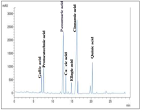
Figure 1: Representative HPLC – MS chromatograph of fractionated phenolic compounds in AECO leaves
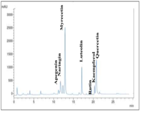
Figure 2: Representative HPLC-MS chromatograph of fractionated flavonoid compounds in AECO leaves
Table 3: Antioxidant activity of AECO leaves against DPPH radical scavenging activity
|
Con. (µg/ml) |
2000 |
1000 |
500 |
250 |
125 |
62.5 |
31.5 |
15.75 |
7.8 |
3.9 |
|
Scavenging activity % |
88.1 |
65.2 |
54.6 |
51.5 |
38.9 |
37.5 |
36.8 |
36.5 |
32.7 |
29.8 |
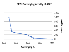
Figure 3. Antioxidant activity of AECO leaves against DPPH radical scavenging
Effect of AECO leaves on the serum levels of hepatic enzymes
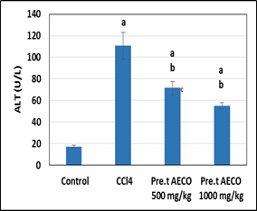
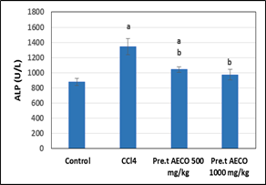
The assay of hepatic functions is an important issue in diagnosis of liver toxicity. The two transaminases enzymes, AST and ALT are shown to be a credible indicator of direct liver cell injury caused by toxic materials including CCl4 [42]. Increased level of ALP is attributed to increased biliary pressure [43]. Liver enzymes rising is a signal of cellular leakage and loss of hepatic functional integrity [42].
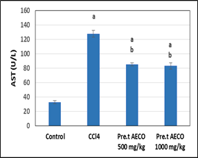
In the current study, the data demonstrated that the rats intoxicated by CCl4 (control positive) for 2 weeks showed a highly significant (sig) (P < 0.05) increased in levels of hepatic enzymes AST, ALT and ALP by 292.02%, 554.4% and 52.6% respectively compared to the normal control as shown in Table 4 and Fig 4, 5 and 6. The raises values of liver enzymes for this group indicated the harmful effect on liver function enzymes. Works done by [13] showed similar effect of CCl4 on liver enzymes, regarding rats injected i.p. with CCl4 (0.2 mL/100 g), mixed with corn oil for 14 days. The increased of hepatic enzymes levels are direct indication of alterations in the liver integrity [44].
In addition, the pretreated rats with low and high doses of AECO had a high sig decrease (P < 0.001) in serum level of AST and ALP enzymes by 33.3%, 34.9%, 18.97%, 27.5% respectively when compared to toxic control. Meanwhile, the serum levels of ALT enzyme in pretreated rats with AECO at (500 and 1000 mg/kg/b.w) and intoxicated with CCl4 recorded a high sig (P < 0.001) decreased by 35.4% and 50.4% respectively compared with the positive control group. At the same time, there was a slightly sig decreasd in ALT between low dose and high dose of protection groups, thus indicating that pretreated rats with AECO (high dose) had a higher reduction of ALT enzyme than those of low dose extract. These findings were consistent with the findings of [5, 20]. They suggested that the ability of extracts to act as protective and curative supplement to prevent the leakage of liver enzymes. This may be due to the presence of antioxidant constituents and polyphenols such as vitamin C, gallic acid and quercetin which act as a free radical's scavenger and responsible for this hepatoprotective activity [45-47].
Table 4: Effect of AECO leaves on the serum levels of liver function enzymes
|
Groups |
AST (U/L) |
ALT (U/L) |
ALP (U/L) |
|
Control negative |
32.6 ± 2.2 |
16.9 ± 1.4 |
881 ± 46 |
|
Control Positive (CCl4) |
127.8 a*** ± 4.8 |
110.6 a*** ± 12.3 |
1344 a*** ± 103 |
|
AECO 500 mg + CCl4 (Pre.t) |
85.2 a***, b*** ± 2.1 |
71.4 a***, b*** ± 6.2 |
1089 a* b** ± 23 |
|
AECO 1000 mg +CCl4 (Pre.t) |
83.1 a***, b*** ± 4.4 |
54.9 a***, b***, c* ± 3.0 |
975 b*** ± 67 |
Results are expressed as mean ± SE (n = 6). a sig versus control group, b sig versus CCl4 group and c low versus high dose treated. (*P < 0.05, ** P < 0.01 and ***P < 0.001).
a sig versus control group, b sig versus CCl4 group and c low versus high dose treated.
Figure 4: Effect of AECO leaves on the serum levels of (AST).
Effect of AECO on the serum levels of malondialdehyde level (MDA) and glutathione peroxidase (GPx)
Lipid peroxidation (LP) has been supposed as the mechanism by which free radical- caused tissue damage and hence, free radical scavenging is established as a mean by which phytoconstituent inhibit LP [48]. Lipid peroxidation is a main sign of hepatotoxicity produced by CCl4 [49]. Liver damage by CCl4 produced from its conversion by the P450 system to the reactive peroxy trichloromethyl radical (OOCCl3) and trichloromethyl radical (CCl3) [12]. These free radicals lead to LP, increase of liver enzymes and reduction of antioxidants capacity [13].
a sig versus control group, b sig versus CCl4 group and c low versus high dose treated.
Figure 5: Effect of AECO leaves on the serum levels of (ALT)
a sig versus control group, b sig versus CCl4 group and c low versus high dose treated.
Figure 6: Effect of AECO leaves on the serum levels of (ALP)
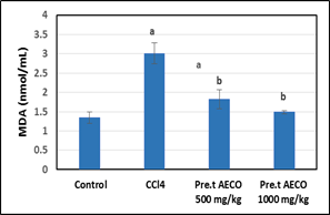
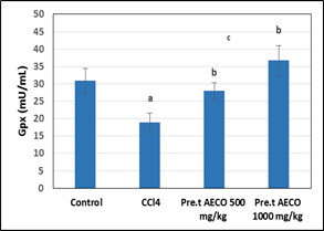
In the present study, the results revealed that the toxic group showed a highly significant increase in serum levels of MDA by 130% and sig decrease in serum levels of GPx by 38.7% compared to the normal control (Table 5 and Figs 7 and 8).
Similar findings were reported by [13, 39, 50] who referred that the increase in MDA levels proves the occurrence of LP, that further results in tissue damage. This could be attributed to damage of the inability of antioxidant defense (GPx) process and to prevent the free radical's generation.
Furthermore, the data ascertained that rats pretreated with AECO (low & high doses) recorded a highly sig decrease in MDA levels by 45% and 54%, respectively compared to CCl4 group. However, more significant effect was observed in rats pre-treated with (1000 mg/kg) compared to low dose group (Table 4.4 and Fig 4.7). Some previous studies have indicated the ability of AECO to decrease the level of LP in rats [20, 37]. They attributed the reduction levels of MDA to the scavenging radicals pointing that the extract possesses a potent antioxidant activity.
Meanwhile, pre-treatment groups, low and high dose group showed a significant (P < 0.05) increase in serum level of GPx by 47.4% and (94.7%), compared with the positive control group. Thus, more significant effect was observed in high dose compared to low dose group. These findings were in agreement with those reported by [20, 37]. Thus, the increase in this antioxidant marker could be attributed to the ability of AECO to stimulate antioxidant enzyme to fight the free radical produced by CCl4.
Table 5: Effect of AECO on the serum levels of MDA and GPx
|
Parameter Groups |
MDA (nmol/mL) |
GPx (mU/mL) |
|
Control negative |
1.3 ± 1.4 |
31 ± 3.7 |
|
Control Positive (CCl4) |
3.3 a*** ± 0.27 |
19 a** ± 2.8 |
|
AECO 500 mg + CCl4 (Pre.t) |
1.8 a* b*** ± 0.25 |
28 b* ± 2.4 |
|
AECO 1000 mg +CCl4 (Pre.t) |
1.5 b*** ± 0.05 |
37 b*** c* ± 4.4 |
The results are expressed as mean ± SE (n = 6). a sig versus control group, b sig versus CCl4 group and c low versus high dose treated. (*P < 0.05, ** P < 0.01 and ***P < 0.001).
a sig versus control group, b sig versus CCl4 group and c low versus high dose treated
Figure 7: Effect of AECO leaves on the serum levels of MDA
Histopathological findings
Compared to the normal liver histology (G1) where liver lobulation looked normal and hepatocytes showed normal cytoplasmic and nuclear features (Fig 9 B); carbon tetrachloride (G2) results in evident hepatocyte necrosis or liver fibrosis depending on dose and duration [6]. Massive swelling, vacuolation and degenerative changes of hepatocytes, inflammatory cell infiltrate at necrotic hepatocytes areas could be observed (stars) (Fig 10 A). Histological finding goes in hand with biochemical increase of liver enzymes due to tissue damage and explained the reported decrease in the protective antioxidant elements. Previous studies using this model, reported similar histopathological alteration [13, 14].
a sig versus control group, b sig versus CCl4 group and c low versus high dose treated
Figure 8: Effect of AECO on the serum levels of and glutathione peroxidase (GPx)
Both low and high doses given prior to CCl4 administration result in sig preservation of normal hepatic structure at both central and portal vein region. In (G3) low dose (500 mg/kg) protection group, potential protection against CCl4 hepatotoxicity was observed where hepatocytes looked normal expects of narrow necrotic zones around the central veins or as scattered few focal necrotic areas among normal tissue (Fig 11 A & B). On the other hand, in (G4) high dose (1000 mg/kg protection group, more protection against CCl4 hepatotoxicity compared to low dose group was observed. There was marked absence of necrotic changes. Hepatocytes and blood sinusoids looked more or less similar to those of the control (Fig 11 C & D). As mentioned before for molokheya (Corchorus olitorius L) as a nutritious food, its leaves are rich in beta-carotene, chlorophylls, phenols, dietary fibers and proteins [51-53]. It could be suggested that such valuable antioxidants constituents are the underlying cause of observed protection or treatment and reduced the risk of liver injury or dysfunction by mitigating hepatic oxidative. Hepatoprotection activity of AECO was observed against other hepatotoxic substances such as lead, cadmium and arsenate and most of such literature attributed their effect to its antioxidant activity [19, 20, 37].
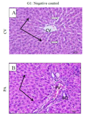
Figure 9: showing normal hepatocytes are arranged in radial plates around the central vein (CV), they have uniform shapes and lightly stain active nuclei (picture A). While picture B showing normal hepatic artery and bile duct (BD). Pv (portal vein)
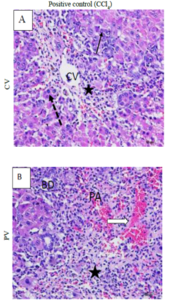
Figure 10: showing inflammatory cells (dotted arrows), massive swelling, vacuolation and degenerative changes of hepatocytes (black arrows). Inflammatory cell infiltrate at necrotic hepatocytes areas could be observed (stars) (A&B).
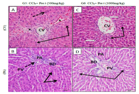
Figure 11: Potential protection was offered by low dose (A&B) against CCl4 hepatotoxicity; hepatocytes ( black arrows) looked normal expects of narrow necrotic zones (stars) around the central veins (CV) or portal veins (PV) in (A&B). while G6 (high dose) showed more protection compared to G5 with marked absence of necrotic changes. Hepatocytes (black arrow) and portal vessels (dotted arrow PV) looked more or less similar to those of control in picture (C&D).
CONCLUSION:
It could be suggested that the AECO leaves have considerable hepatoprotective effect against CCl4 induced liver damages by suppressing oxidative injury in the livers of rats and reduction in the morphological changes caused by toxin. The antihepatotoxic activity of AECO leaves could be attributed to their radical scavenging activity because they are rich in flavonoids and phenolic compounds.
REFERENCES


