International Journal of Pharmaceutical and Phytopharmacological Research
ISSN (Print): 2250-1029
ISSN (Online): 2249-6084

The purpose of this research was to estimate the hepatoprotective efficacy of clay nanoparticles in improving the effects of anticancer drugs such as doxorubicin (DOX). One hundred male adult mice were randomly divided into five groups. Group 1 (control) was injected with double distilled water, Group 2 (EAC group) received intraperitoneal (IP) injection of 0.15 ml Ehrlich cells (2×10⁶), in Group 3 (EAC+DOX group) EAC-bearing mice were treated with 0.07 ml of doxorubicin at a dose of 10 mg/kg bw. Doxorubicin was administered IP in six equal doses of injections to animals for 2 weeks for an accumulative dose of 10 mg/kg bw. Group 4 (EAC+DOX+MMT group) got i.p. infusion of 0.07 ml doxorubicin (10mg/kg) stacked on Montmorillonite nanoparticles (30 mg/kg) 3 times per week for about fourteen days. Group 5 (EAC+DOX+OCTA+MMT group) was gotten i.p. administration of 0.07 ml doxorubicin (10mg/kg) stacked on Octadecylamine (OCTA) with Montmorillonite nanoparticles (30 mg/kg) 3 times each week for about fourteen days. Blood samples were obtained at the end of the study and the serum was separated to measure liver function. The liver was removed and histologically analyzed. The serum levels of gamma-glutamyl transferase (GGT), alkaline phosphatase (ALP), alanine aminotransferase (ALT), and aspartate aminotransferase (AST) were elevated, whereas the total protein and albumin were lower than control and other groups in the EAC. After handling of DOX clay NPs, the levels of these parameters were enhanced. In conclusion, nanoclays are influential in curing Ehrlich-induced ascites carcinoma in mice models in the Doxorubicin delivery system. They target DOX release in cancer cells and diminish the side effects of DOX in the liver.
INTRODUCTION
Ehrlich ascites carcinoma (EAC) is a type of homogenous carcinoma, which is established with rapid proliferation, high transplant capacity, and small duration [1]. EAC resembles tumors in humans; therefore, solid and ascetic forms of carcinoma are often used to estimate the anticancer efficiency of different materials [2, 3]. Doxorubicin and adriamycin, among others, are a chemotherapy solution used for cancer therapy [4, 5]. It is used together with other chemotherapy agents [6]. Doxorubicin is administered by infusion into a vein [7, 8]. Doxorubicin binds to nucleic acids by intercalating the double DNA helix and balancing the excision buildings of topoisomerase II, which causes the DNA chain to break at particular destinations prompted by doxorubicin [9]. It works to some extent by interfering with the ability of DNA [10]. The clinical use of DOX is related to the danger of enlarged cardiomyopathy or congestive heart attack [11]. Until now, many kinds of drug-release nano-systems have been created, for example, poly (D, L-lactide- coglycolide) nanopolymers (PLGA), micelles, silica, dendrimers, liposome nanoparticles [12]. Much attention is paid to investigate the scope of drug delivery by using particle delivery systems, including mails for large and small molecules. Particle systems, including nanoparticles, have been successfully used as a physical method to also modify, enhance the pharmacodynamic and pharmacokinetic characteristics of different types of medicinal molecules [13]. Although the supply of anti-cancer corrective operators to strong tumors remains complicated, nanoparticles are an attractive drug delivery system, since they obviously affect cancer cells and have limited damage to typical tissues [14]. In the application of pharmaceutical products, the nano-clay is a part of the area of birth age, since the delivery of the drug is concerned with the release of a controlled drug. Nano-clays have significant potential compared to polymer and the study of drug-clay interactions for the preparation of clay-based drug delivery systems [15]. The famous montmorillonite (smectite) (MMT) nano-clay plate formed by aluminosilicate slabs of approximately 1 nm of surface replaced by metal cations and organized in multi-layer reserves of approximately 10 µm [16]. As far as we know, there are some previous studies in the literature on the influence of clay nanoparticles on the activity of the drug versus tumor. Doxorubicin used for eradication of experimentally induced carcinoma. Therefore, this study was planned to elaborate on the possible hepatoprotective role of clay nanoparticles on the efficacy of doxorubicin in Ehrlich ascites carcinoma in adult male mice.
MATERIALS AND METHODS
Chemicals
Hydrophilic Montmorillonite with Nanoclay (682659, Sigma- Aldrich, Germany); Surface modified MMT Nanoclay containing 25-30 wt. % Octadecylamine (682616, Sigma-Aldrich, Germany), Biochemical kits; Doxorubicin (25316-40-9, Sigma-Aldrich, Germany).
TEM examination
In this study, the MMT NPs were identified by using transmission electron microscopy (TEM). Using a sonicator (BRANSON 1510), MMT and OCTA-MMT Nano-powders were dissolved in 70% ethyl alcohol solution. Then, the suspended MMT and octa- MMT NPs were mounted on a coated carbon copper grid. The morphology of samples was examined by transmission electron microscopy (TEM; JEM-1011, Jeol, Tokyo, Japan) instrument operating at an accelerating voltage of 100 KV [17].
Experimental design
One hundred adult male Swiss mice, weighing (22- 25 grams) were used in this experimental study. These animals were supplied from the animal house of King Fahd Medical Research Center, King Abdulaziz University, Jeddah, Saudi Arabia. All investigation steps were carried out as indicated by Canadian Ethical standards for trial creatures use. Endorsement of the examination configuration was gotten from the Local Biomedical Ethical Committee of King Abdulaziz University, Jeddah, Saudi Arabia. Mice were put in plastic enclosures (20 mice/group) and kept in controlled lab conditions at room temperature (20±1Cº), light: dark cycle of 12:12h, and humidity of (65%), and fed ad libitum with a standard diet containing 11.7% fat calories (20% soybean, 8% concentrated proteins, 50% wheat, 21% corn, and 1% vitamins & salts) and had free intake to tap water. After 1 week, they were randomly divided into 5 groups (20 mice each). Group 1 (control) was injected with double distilled water, Group 2 (EAC group) received intraperitoneal (IP) injection of 0.15 ml Ehrlich cells (2×10⁶), in Group 3 (EAC+DOX group) EAC-bearing mice were treated with 0.07 ml of doxorubicin at a dose of 10 mg/kg bw. Doxorubicin was administered IP in six equal doses of injections to animals for 2 weeks for an accumulative dose of 10 mg/kg bw in line with Li and Singal [18]. Group 4 (EAC+DOX+MMT group) got i.p. infusion of 0.07 ml doxorubicin (10mg/kg) stacked on Montmorillonite nanoparticles (30 mg/kg) 3 times per week for about fourteen days. Group 5 (EAC+DOX+OCTA+MMT group) was gotten i.p. administration of 0.07 ml doxorubicin (10mg/kg) stacked on Octadecylamine (OCTA) with Montmorillonite nanoparticles (30 mg/kg) 3 times each week for about fourteen days.
Blood sampling
Blood samples were gathered inside 24 days post intraperitoneal vaccination of Ehrlich cells [19]. Blood was assembled from retro‐orbital venous plexus into plain sterile content cylinders without anticoagulants. The blood samples were centrifuged at 3000 rpm for 10min and blood sera were gathered, aliquoted, and kept at -80 °C till use [20].
Liver function test
Alanine aminotransferase (ALT) (REF.K2143, Siemens Com., Munich, Germany): The Dimension Vista ALT technique as suggested alanine aminotransferase method of the IFCC as mentioned by [21]. The method depends on the steps outlined by [22]. Alanine aminotransferase activity was estimated utilizing a bichromatic (340, 700 nm) rate method. Aspartate aminotransferase (AST) (REF. K2041, Siemens Com., Munich, Germany): AST levels can be elevated even before clinical jaundice appears [23]. The AST activity was estimated by using a bichromatic (340.700 nm) rate method. Alkaline phosphatase (ALP) (REF. K2115, Siemens Com., Munich, Germany: ALP method is based on the primary reference process for estimation of alkaline phosphatase catalytic activity at 37°C as mentioned by the International Federation of Clinical Chemistry. Alkaline phosphatase technique depends on the method made by [24] and modified by [25]. Alkaline phosphatase was estimated by using bichromatic (405, 700 nm) rate method. Total proteins (TP) (REF. K1073, Siemens Com., Munich, Germany): Method of measurement of total protein was modulation of biuret reaction first presented by [26] and later modified by [27] and presented as the method of choice for serum by [27]. Gama-glutamyl transferase (GGT) was determined by the colorimetric method (rate method). The substrate of this enzyme is L-gamma-glutamyl-3-carboxy-4-nitroanilide transfer to glycylglycine. The amount of 5-amino-2-nitrobenzoate liberated is proportional to the GGT activity and can be photometrically determined [28, 29]. The total protein contents were assessed using a bichromatic (540 & 700 nm) endpoint technique. Albumin (ALB) was used as a reference. The method of albumin measurement is an adaptation of the bromocresol purple dye-binding methods as mentioned by [30, 31]. The complex was absorbed at 600 nm and it was estimated utilizing a polychromatic endpoint method (600, 540, 70 nm).
Histopathology examination
At end of the experimental period, mice were anesthetized with diethyl ether. Then, the liver was removed, fixed in isotonic saline solution and stored in 10% formalin, and then processed paraffin embedding [32, 33]. Five to seven micrometers of thick sections were stained with hematoxylin and Eosin according to [34] and inspected under Olympus trinocular microscope (BX-51). The samples’ photomicrographs were taken under different magnification powers.
Statistical analysis
The qualities were measured as mean ± standard deviation (SD) and SPSS version 23 was utilized for examination (IBM SPSS, IBM Corp., Armonk, N.Y., USA). Factual examinations were made by a One-Way investigation of fluctuation (ANOVA). P-values of <0.05 were considered statistically significant.
RESULTS AND DISCUSSION
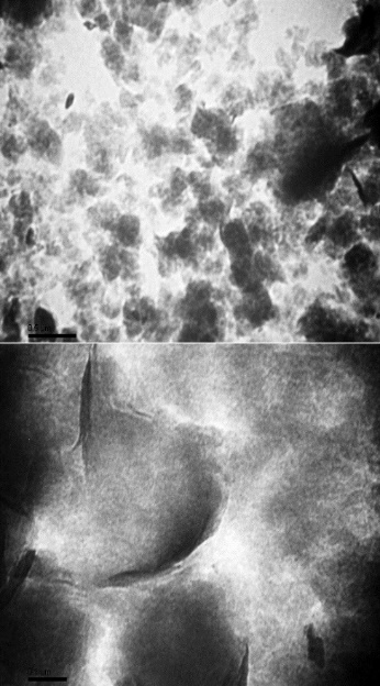
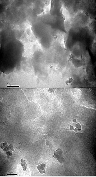
The surface morphology of the Montmorillonite Nanoclay was investigated using TEM, and the micrographs are presented in Figure 1. According to the figure, the Montmorillonite Nanoclay exists in a flaky shape in both exfoliation and intercalation of different layers in nano-forms.
|
|
|
|
a) |
b) |
|
Figure 1. a) TEM micrographs at different magnification power of Hydrophilic Montmorillonite Nanoclay, b) octadecylamine surface modified Montmorillonite Nanoclay |
|
Figure 1 shows a typical TEM micrograph for octadecylamine surface-modified Montmorillonite Nanoclay. The ordered sheets of the nanoclay observed in the TEM image are strong evidence of the intercalated structure, whereas the disordered sheets are evidence of the exfoliated structures octadecylamine surface-modified Montmorillonite Nanoclay and there are large aggregates and agglomerates of Octa/MMT composites.
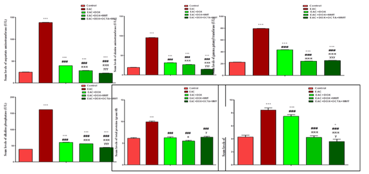
The serum levels of ALP, GGT, AST, and ALT were raised in EAC, EAC+DOX, EAC+DOX+MMT, and EAC+DOX+OCT+MMT groups (P=0.0001 for all) versus control group. The ALT, AST, ALP and GGT levels were significantly diminished in EAC+DOX, EAC+DOX+MMT and EAC+DOX+OCT+MMT groups versus EAC group (P=0.0001 for all); in EAC+DOX+MMT and EAC+DOX+OCT+MMT groups versus EAC+DOX (P=0.0001 for all) and in EAC+DOX+MMT versus EAC+DOX+OCT+MMT groups (P =0.0001, P =0.0001, P=0.0001 and P=0.005, respectively). Serum bilirubin levels were significantly higher in EAC and EAC+DOX groups versus control (P=0.006 and P=0.014), however, was significantly lower in the EAC+DOX+OCT+MMT group versus EAC, EAC+DOX, and EAC+DOX+MMT (P=0.001, P=0.002, and P=0.029, respectively) (Table 1 and Figure 2).
All serum levels were significantly lower in EAC and EAC+DOX groups than control (P=0.0001 for both). In the meantime, absolute protein levels raised significantly in EAC+DOX+MMT and EAC+DOX+OCT+MMT groups versus EAC+DOX (P=0.008 and P=0.0001, respectively). Serum albumin levels were significantly declined in EAC than control, EAC+DOX, EAC+DOX+ MMT and EAC+DOX+ OCT+MMT groups (P=0.005, P=0.015, P=0.002, and P=0.010, respectively); in EAC+DOX+ OCT+MMT versus EAC+DOX and EAC+DOX+MMT (P=0.034 and P=0.005, respectively) (Table 1 and Figure 2).
Table 1. Comparison of liver function test in different study groups
|
Groups Parameters |
Control |
EAC |
EAC+DOX |
EAC+DOX+MMT |
EAC+DOX+OCTA+ MMT |
|
ALT (U/L) |
19.32±0.35 |
95.50±0.50 |
31.28±0.30 |
26.42±0.38 |
14.42±0.38 |
|
Significance |
- |
1P =0.0001 |
1P=0.0001; 2P=0.0001 |
1P=0.0001 ;2P=0.0001, 3P=0.0001 |
1P=0.0001; 2P=0.0001, 3P=0.0001; 4P=0.0001 |
|
AST (U/L) |
24.93±0.71 |
138.25±0.25 |
39.42±0.38 |
28.33±0.38 |
22.25±0.25 |
|
Significance |
- |
1P =0.0001 |
1P=0.0001; 2P=0.0001 |
1P=0.0001; 2P=0.0001, 3P=0.0001 |
1P=0.0001; 2P=0.0001, 3P=0.0001; 4P=0.0001 |
|
ALP (U/L) |
39.25±0.25 |
161.42±0.38 |
60.42±0.38 |
56.25±0.25 |
44.42±0.38 |
|
Significance |
- |
1P =0.0001 |
1P=0.0001; 2P=0.0001 |
1P=0.0001; 2P=0.0001, 3P=0.0001 |
1P=0.0001; 2P=0.0001, 3P=0.0001; 4P=0.0001 |
|
GGT (U/L) |
22.47±0.42 |
79.43±0.40 |
43.40±0.36 |
24.25±0.25 |
25.33±0.38 |
|
Significance |
- |
1P =0.0001 |
1P=0.0001; 2P=0.0001 |
1P=0.0001; 2P=0.0001, 3P=0.0001 |
1P=0.0001; 2P=0.0001, 3P=0.0001; 4P=0.005 |
|
Total protein (gram/dl) |
6.12±0.63 |
4.30±0.18 |
4.52±0.25 |
5.50±0.30 |
6.08±0.33 |
|
Significance |
- |
1P =0.0001 |
1P=0.0001; 2P=0.522 |
1P=0.068; 2P=0.003, 3P=0.008 |
1P=0.914; 2P=0.0001, 3P=0.0001; 4P=0.082 |
|
Albumin (gram/dl) |
4.28±0.30 |
3.48±0.31 |
4.12±0.13 |
4.38±0.13 |
3.58±0.38 |
|
Significance |
- |
1P =0.005 |
1P=0.483; 2P=0.015 |
1P=0.658; 2P=0.002, 3P=0.264 |
1P=0.010; 2P=0.658, 3P=0.034; 4P=0.005 |
Data were communicated as mean ± standard deviation. 1P: Significant change contrasted with the benchmark group; 2P: Significant change contrasted with the Ehrlich Solid Carcinoma (EAC) group; 3P: Significant change contrasted with the Ehrlich Solid Carcinoma and DOX (EAC+DOX) group; 4P: Significant change contrasted with the Ehrlich Solid Carcinoma +DOX+MMT (EAC+DOX+MMT) group. Hugeness was made utilizing ANOVA-one path test at P ˂0.05. ALT: alanine aminotransferase; AST: aspartate aminotransferase; ALP: alkaline phosphatase; GGT: gamma-glutamyl transferase.
|
|
|
Figure 2. Comparison of liver function tests in different study groups |
Histological changes in liver
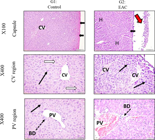
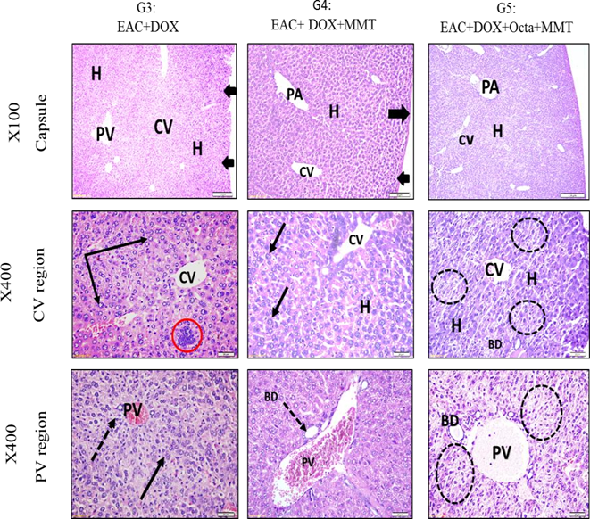
Histological study was combined with the biochemical investigation to confirm the possible hepatoprotective effect of clay nanoparticles. Control liver (G1) showed normal lobulation extended between central veins and portal areas. Hepatocytes showed acidophilic cytoplasm and active one or two nuclei. Hepatocyte cords were separated by normal blood sinusoids. Portal regions showed normal contents (branches from the portal vein, bile duct, and hepatic artery) and normal nearby hepatocytes (Figure 3). EAC (G2) showed aggregation of Ehrlich cells near the capsule. Numerous hepatocytes looked shrunken with dark cytoplasm and small degenerated. Pyknotic nuclei. Blood vessels showed congestion. The Portal area showed a congested portal vein. Nearby hepatocytes also showed similar degenerative changes. Such changes indicated the affection of liver tissue by the presence of EAC cancer cells near their capsule or even invading liver tissue (Figure 3). On the other hand, EAC+DOX (G3) where DOX, was given to mice–bearing EAC resulted in decreased migration of tumor cells from the peritoneal cavity to nearby-organs. High power showed liver tissue with the absence of tumor cells near the capsule. Normal hepatocytes had central rounded nuclei similar to control. Other hepatocytes either showed abnormal large nuclei or nuclei with chromatin granules. Few regions showed aggregated degenerated hepatocytes (Figure 4). EAC+DOX+MMT (G4) showed normal hepatocytes, which looked healthy with round vesicular nuclei. Blood sinusoids between cells were normal. Some cells showed large nuclei (karyomeglay). The portal area region showed also no tumors, and hepatocytes looked normal with no signs of degeneration or necrosis. Portal vein showed dilatation and congestion (Figure 4). EAC+ DOX+ OCTA+ MMT (G5) showed no hepatic changes. Hepatocytes showed enlarged nuclei (karyomeglay) with increased chromatin granules. Central veins and portal vessels showed a normal appearance of blood vessels. It also showed the absence of tumor cells near the capsule or inside the liver parenchyma. Hepatocyte looked more or less normal with round vesicular nuclei. Some samples still showed foci of degenerative changes similar to those seen in liver mice treated with DOX.
|
|
|
Figure 3. Sections from mice liver stained by Hematoxylin and Eosin and photographed at different powers X100 & X400 (G1- G2) |
|
|
|
Figure 4. Sections from mice liver stained by Hematoxylin and eosin and photographed at different powers X100 & X400 (G3-G4- G5) |
The TEM images represented an exfoliated/intercalated nanocomposite. This is probably because of the organic modification of MMT that provides the possibility for octadecylamine chains to diffuse between the layers during processing [35].
Key problems accompanied by cancer therapies are severe side effects that result from normal tissue destruction, including heart, liver, and spleen [36-40]. The hepatotoxic actions of anticancer drugs may manifest as symptoms other than liver injury, as necrosis, fibrosis, steatosis, cholestasis, and vascular injury [41]. Hao et al. [42] revealed that nanoparticles could decline DOX toxicity, which results in an elevation of the average lifetime compared with free DOX. During malignancy, enzymatic alterations of tumor markers reflect on the digestion settings, and they are identified as the basis for ensuring disease cell behavior with affectability and particularity [43]. The antitumor action of Doxorubicin is primarily interceded for numerous reasons such as genomic DNA, cell-cycle arrest, and apoptosis. In addition, doxorubicin-induced reactive oxygen species drive their toxicity through redox cycling process [44]. Dimitrakis et al. [44] tested the hypothesis that anthracyclines affect protein degradation pathways in adult cardiomyocytes connected to the impacts of autophagy, apoptosis, and proteasome/ubiquitin framework in long-term cultured adult rat cardiomyocytes. In clinical applications, regardless of doxorubicin dose-dependent cardiotoxic effects, it remains in use because of its usefulness in a many kinds of tumors treatment [45]. Drugs can be encapsulated in particles or added to the surfaces of the nanoparticles. The basis for increased tumor specificity is the differential accumulation of drug-loaded nanoparticles in tumor tissue versus normal cells, which results from particle size rather than binding. Recently, coordinated therapeutics in nanomedicine has been explored in most cases [46].
The liver is the largest vital organ that plays an important role in many functions in the body, such as the synthesis of blood clotting factors, proteins, triglycerides, cholesterol, glycogen, and bile. Serum liver enzymes are used for the early prognosis of neoplasm, metastasis, and malignancy [47]. The liver diseases have become an important cause of mortality throughout the world. Among them, chemical-induced injury is one of the common causative factors that pose major clinical and regulatory challenges. Liver injury has become a serious health problem due to the use of many prescription drugs and exposure to various toxins [48]. Liver index is a precise way to deal with decide adjustment in liver size contrasted with the estimation of the liver weight alone as the liver weight chiefly relies upon liver size [49].
This study showed increased serum levels of AST, ALT, ALP, GGT, and decreased total protein and albumin in EAC-treated group compared to the control. Moreover, the level of these parameters decreased after DOX treatment in the EAC+DOX+ MMT and EAC+DOX+OCTA+MMT groups. AST enzyme is employed in the evaluation of hepatic disorders and an increase in this enzyme activity reflects acute liver damage and hepatocellular disorders. ALT is an important biomarker for liver toxicity. ALT is increasingly associated with liver injury compared to AST. These enzymes are cytoplasmic in nature, but in the event of liver damage, they enter the circulatory system due to changes in the permeability of the hepatocyte membrane [50]. Mesalam [51] demonstrated that liver ALT and AST increase in EAC-bearing mice compared with the control mice. Damaged hepatocytes in the bearing-mice with EAC cells lead to the diffusion of these intracellular enzymes into the circulation [52]. This can increase the ALT and AST levels in the serum of mice with EAC [53].
EAC infused in mice significantly raised serum levels of ALT, AST, and total bilirubin though levels of serum albumin and total protein declined significantly compared to the control mice, suggesting compromised liver function [54]. The total protein levels are decreased in hepatotoxic conditions as consequences of modification in the protein, lipid, carbohydrate or perturbed protein biosynthesis in the cirrhotic liver [55]. Increased serum bilirubin levels might be due to bile ductile, inflammation and fibrosis in portal triads and/or regurgitation of conjugated bilirubin from necrotic hepatocytes to sinusoids [56]. Increased serum ALP is a direct result of its increased synthesis, in the presence of increasing biliary pressure [57]. Our results were confirmed by changes in the liver histology, which demonstrated necrosis, degeneration, appoptosis, and fibrosis. Tohamy et al. [58] found that after 10 days of EAC inoculation, there were regular reductions of hepatic total bilirubin levels in the liver of EAC-bearing mice in comparison to the normal group.
Dolai et al. [59] reported that EAC mice show increased activities of liver markers such as ALT, AST, due to hepatocellular damages. The liver metabolic changes might be due to raised levels of decarboxylase stimulating parameters, tumor necrosis factor (TNF-α), and interleukin-1 (IL-1) synthesized by tumor cells [60]. A huge body of evidence suggests that reduced total protein levels are a key factor in the improvement of liver damage [61]. Junior et al. [62] showed that the level of albumin significantly decreased in the EAC-bearing mice.
Mesalam [51] showed a normal hepatic architecture with no specific histopathological lesions in the control group while liver sections of mice bearing Ehrlich ascites carcinoma (EAC) noted various histopathological alternations including, vacuolization of hepatocellular cytoplasm, and sporadic necrosis of hepatocytes with deeply pyknotic nuclei, congestion of central vein associated with brown pigment deposition, and area of hemorrhage. Liver tissues revealed minimal infiltration with no fatty generation, or nodule formation or dilatation of central vein, or necrosis in normal mice while many organs of EAC-bearing mice revealed marked cellular degeneration/regeneration as results of carcinogenesis [63].
The histopathological alterations noticed in the doxorubicin only group were the same as those observed previously [64-66]. Histopathology examination of liver in group 3 (EAC+DOX) indicated sinusoidal blockage and mononuclear cells invasion, however, aggregated/degenerated hepatocytes were lower in the EAC group. There was no corruption, metastatic foci or EAC attack in the liver while in group 4 (EAC+DOX+MMT), there was a mild sinusoidal blockage, moderate vacuolation, and mononuclear cell penetration, yet seriousness was little compared to the EAC group [54]. Mice treated with DOX showed degeneration of hepatocytes with pyknosis and karyolysis of their nuclei. Focal vein dilatation with drain, monocytes invasion, and enlarged blood sinusoids was observed [67].
CONCLUSION
Clay nanoparticles are effective in DOX delivery system for the treatment of Ehrlich-induced ascites carcinoma in mice model. They targeted DOX release to the cancer cells and minimized the side effect of DOX in the liver.
Acknowledgments: None
Conflict of interest: None
Financial support: None
Ethics statement: None