International Journal of Pharmaceutical and Phytopharmacological Research
ISSN (Print): 2250-1029
ISSN (Online): 2249-6084
|
Studying the Histopathological Effects of Aqueous Extract of Cola nitida on Adult Rats’ Liver
Laila A Hummdi*, Waffa A Zabarmawi, Abeer M Waggas |
|
Department of Biology, University of Jeddah, Jeddah, Saudi Arabia. |
|
ABSTRACT |
|||||||||||||||||||||||||||||||||||||||||||||||||||||||||||||
|
The present work aimed to study the effects of daily use of Kola nut on liver tissue. The experiment was carried out on 40 male Wister albino rats, which were divided into 4 groups. The first group (G1) served as a control group. Groups G2 and G3 were treated with 10 mg/Kg body weight of aqueous Kola nut extract for 15 and 60 days, respectively. The rats in Group G4 were treated with 10 mg/Kg body weight of aqueous extract of Kola nut for 60 days, then they were left for normal recovery without treatment for 30 days. The rats were sacrificed and their liver tissues from different groups were processed and examined after 15 and 60 days and the withdrawal group after 30 days of stopping the treatment. The histological results showed that the treatment for 15 days caused acute congestion of the liver tissues, dilated central veins with erythrocytes stasis, edematous, inflammatory cell infiltration, and lack in Kupffer cell number. The liver of rats treated for 60 days represented fatty infiltration, vacuolar cytoplasmic degenerations, cellular necrosis, nuclear karyolysis, and bile duct proliferation. While the liver treated for 60 days then left for normal healing for 30 days showed continuous histopathological damage as well as severe disorganization of normal liver tissue structure with a significant decrease (P <0.05) in relative liver weight. The results of the current study indicated that aqueous extract of Kola nut induced hepatotoxicity via oxidative stress. |
|||||||||||||||||||||||||||||||||||||||||||||||||||||||||||||
|
Key Words: Cola nitida, hepatotoxicity, rats, histopathological changes. INTRODUCTION The liver is one of the most vital organs [1-3] in the body as it regulates all metabolic processes [4] and performs many vital functions as it plays a role in the metabolism of fats, carbohydrates, proteins, detoxification, and immunity, as well as the production and secretion of bile, albumin, clotting factors, cholesterol and steroid hormones. Although the liver is not directly connected to pollutants, it is indirectly affected due to its direct contact with blood, which exposes it to pathological changes and metabolic disorders in many mammals [5-7]. Kola nut has different types, the most common of which are cultivated types in Nigeria: Cola nitida and Cola accuminata [8]. Cola nitida (Kola nut) is a seed of a tropical tree that belongs to the Sterculiaceae family [9, 10]. The aqueous extract of kola nut contains 13.5% water, 9.5% protein, 1.4% fat, 45% sugar, cornflour 7.0%, and cellulose; kola seeds are also rich in caffeine (6.2%), theobromine 0.9%, and catechin 15% [11, 12]. Recently it was found that the kola nut contains kolanin [13]. Several researches stated that caffeine is one of the most important components of Kola nut, which is mainly responsible for its physiological activities [14-17]. The percentage of caffeine may differ from one study to another and the reason may be due to the methods of preparation used and other factors such as the duration of the experimentation period and plant collection process, in addition to geographic location or habitat [18]. The nature of the climate affects the concentrations of active substances, especially alkalis and phenolic compounds present in plants [19-21]. Kola nut is used in many industries for example oil spices, soft drinks, Cola drinks, sweeteners, and in caramel and chocolate [22, 23]. Kola nut has been used in folk medicine as an aphrodisiac, an appetite suppressant, nausea, migraine, indigestion treatment, and in some cases, it is used to control vomiting in pregnant women [24]. The plant was also used in treating skin wounds and infections [25]. However, kola nut extract and caffeine showed toxic effects that may lead to death in rats within 48 hours, and the study also stated that the oral LD50 dosage of Kola nut and caffeine was about 150-200 mg/kg [21]. A study was conducted to determine the effect of Kola nut extract on the total body weight of rats and pointed that it decreased with an increase in the size of the liver, kidneys, brain, and testis [26], with the occurrence of some cellular changes [27]. Other studies recorded a decline in the mobility of mice after chronic consumption of Kola nut, in addition to a reduced intake of food and water that led to bodyweight loss, and attributed that Kola nut may have an appetite suppressant effect [17, 28, 29]. Individuals who almost daily consume Kola nut were sufferers of tumors manifestations [30]. Conversely, the consumption of Kola fruits had a toxic effect on cancer cells in the human liver [31]. It has been confirmed that Kola nut has negative effects on the hepatocytes and blood vessels [32, 33]. A recent study concluded that Kola nut has a toxic effect on kidney and liver functions in rats [34]. Kola nuts are widely available in public markets of Saudi Arabia, as a natural stimulant, and used indiscriminately as an aphrodisiac, anti-fatigue, and for diabetes treatment. The present study is aimed to evaluate the histological effect of the Cola nitida on the liver of male albino rats and to accommodate the scarcity of research on the safety of this plant on liver tissues. MATERIALS AND METHODS Experimental animals This study was conducted on 40male adult albino rats (Rattus norvegicus), with a weight ranged between 150-200 g. They were obtained from the King Fahd Center for Medical Research of King Abdulaziz University in Jeddah. Experiments were conducted in the same center, where the animals were raised in special cages and exposed to suitable environmental temperature conditions of 22-25 °C, humidity ranging from 30% to 72%, good ventilation, and provided with appropriate feeding and water throughout the treatment period. Plants The Kola nut fruits were obtained from a popular local market in Jeddah, Saudi Arabia. Preparation of Cola nitida aqueous extract The aqueous extract was prepared according to a method described previously [35]. 50g of Kola nut was crushed with 100 ml physiological solution. Then, the mixture was placed in a dark-colored glass bottle and left for 24 hours. The floating precipitate of Kola nut was collected from the mixture and filtered. The extract was diluted according to the dosage used in the method [36]. Experimental design 40 male rats were divided into 4 groups, and each group contained 10 rats. In the first group (G1), the rats were injected with a physiological solution as a control group. The rats in the second group (G2) were treated intraperitoneally (ip) with the aqueous extract of kola nut at a dosage of 10 mg/kg according to [35, 36], for a period of 15 days. The rats in the third group (G3) were also treated similarly at a dose of 10 mg/kg for 60 days. In the fourth group (G4) or withdrawal group, the rats were treated ip with aqueous extract of kola nut at a dose of 10 mg/kg for 60 days, then the rats were left without treatment for 30 days. Tissue samples were collected after 30 days of stopping the treatment to study the histopathological effects. Liver tissue samples were taken from G1 and the treated groups to study the histopathological effects after 15 and 60 days respectively of G2 and G3, and after 30 days of stopping treatment in G4.
Histological study At the end of each experimental period, the rats were sacrificed and the liver tissue samples were harvested, rinsed in phosphate-buffered saline, and then fixed in 10% buffered formalin for subsequent histopathological examination. The tissues were processed, embedded in paraffin, sectioned at a thickness of 2µm, and stained with hematoxylin-eosin using a standard method according to [37].
Statistical analysis The body and liver weights changes were measured in rats treated with Kola nut extract compared to the control group using Statistics SPSS 24 software. The changes in the liver weights were expressed as mean and ± SEM using the Student t-test. The percentage difference was also calculated compared to the control group according to the following equation [38, 39].
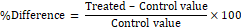
%Difference = Treated – Control valueControl value×100 RESULTS Histological changes in the liver of treated rats Control rats (Fig. 1a, b, c) Liver examination of adult control rats under light microscopy revealed that the liver consists of hepatic lobules intertwined with each other and not separated by barriers of connective tissue stroma characteristic found in mammals. Hepatocytes are arranged in interconnected plates, which are often single-cell polygonal-shaped that contain a central basic nucleus, either one or two nucleolus and acidophilic cytoplasm (Fig. 1a). Hepatocytes arise from central veins and are separated by blood sinusoids that contain two types of cells. Endothelial cells are characterized by oval nuclei and Kupffer cells are characterized by triangular nuclei (Fig. 1a, b). Liver tissue in rats is also characterized by the presence of portal areas (Fig. 1c) that contain a vein, portal artery, and one or more bile ducts lined with an epithelial layer consisting of cuboidal cells containing large vesicular nuclei, as well as connective tissue histiocytes spread in portal areas in small amounts.
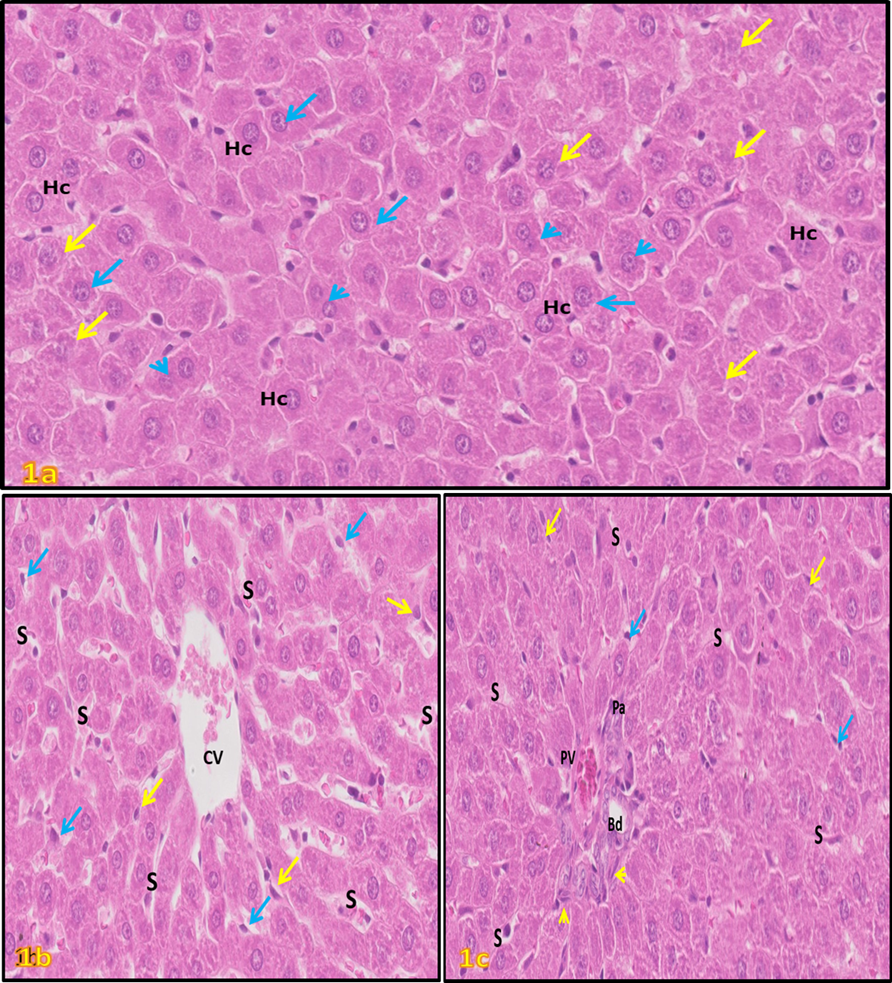
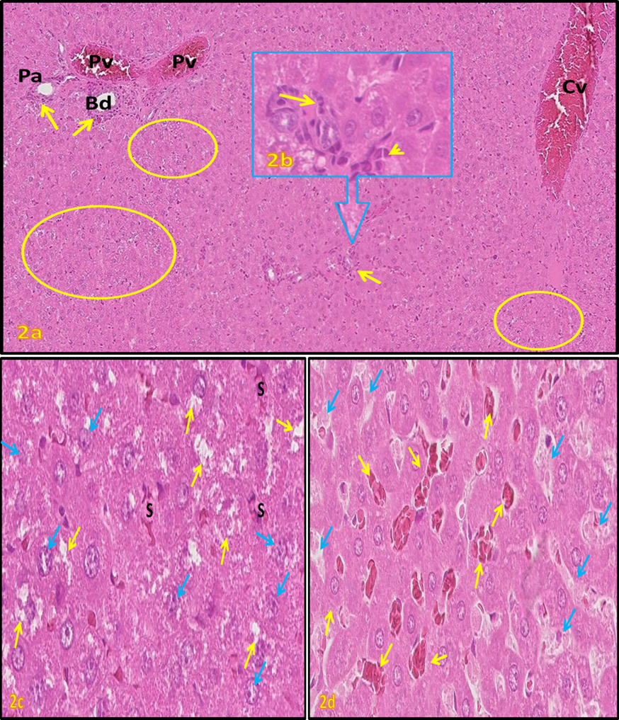
Treated rats (Fig. 2, 3, 4) Liver examination sections of the rat groups treatment for 15 days with aqueous extract of Cola nitida showed an occurrence of vacuolar degeneration of hepatocytes, acute congestion of liver tissues, dilated central veins, blood sinusoids associated with red blood cells stasis, inflammatory cells infiltration, and Kupffer cells loss (Fig. 2a, b, c, d). These previous changes were associated with a significant increase (P <0.05) in the mean relative weight of the liver compared to the control group (Table 1). The liver damage was intensified with an increase in the treatment period to 60 days, represented by the walls of central veins that were damaged and fibroid (Fig. 3a). The degenerative changes in hepatocytes amplified in some sections which showed vacuolar cytoplasmic degeneration, cellular necrosis, and nuclear karyolysis (Fig. 3a, b). However, massive fatty infiltrations (lipid vesicles) were observed in liver tissue (Fig. 3c). Bile duct proliferation, fibrosis of connective tissue around the portal tract, and leukocytes margination to the edge of the vessel's areas were observed (Fig. 3d). These changes were associated with a non-significant increase in the size of hepatocytes and the relative weight of the liver compared to the control group (Table 1). While the liver treated with aqueous extract of cola nut for 60 days then left for normal healing without treatment for 30 days showed continuous histological damage as well as severe disorganization of normal liver tissue structure (Fig.4a, b, c) with a significant decrease (P <0.05) in relative liver weight.
Figure (1a-c): Light micrographs (L.M.) of liver sections of control groups of adult rats; hematoxylin and eosin-stained (H. & E.). (a): normal hepatocytes (Hc) with granular cytoplasm, some hepatocytes with basophilic masses (yellow arrows), round clear nucleus (blue arrows) contains scarce heterochromatin, binucleated cell (arrowheads); H. & E. (400X). (b): hepatic plates radiated from the central vein (CV) and separated by blood sinusoids (S), Kupffer cells (blue arrows), endothelial cells (yellow arrows); H. & E. (400X). (c): portal area; portal vein (Pv), portal artery (Pa), bile ducts (Bd), and histiocytes (arrowheads). Note endothelial cells (yellow arrows), blood sinusoids (S), Kupffer cells (blue arrows); H. & E. (400X).
Figure (2a-d): L.M. of liver sections of adult rats treated with aqueous extract of kola nut with a dose of 10 mg/kg.BW/day for 15 days (H. & E.). (a): Congestion and dilated central (CV), portal veins (PV), portal artery (Pa), mononuclear leukocyte aggregates around the portal area and in hepatic tissue (yellow arrows), vacuolation of hepatocytes (circles); H. & E. (100X). (b): Enlarged part from (2a) section showing mononuclear Leukocyte aggregates containing lymphocytes (arrow), macrophage (arrowhead); H. & E. (1000X). (c): Vacuolated hepatocytes with cytoplasmic degeneration (yellow arrows), pleomorphic nuclei (blue arrows), sinusoids (S) congestion; H. & E. (400X). (d): Blood sinusoids disorganized. Note congestion (yellow arrows), dilated sinusoids lumen (blue arrows) and filled with cellular debris, loss of Kupffer, and endothelial cells; H. & E. (400X).
Figure (3a-d): L.M. of liver sections of adult rats treated with aqueous extract of Kola nut with a dose of 10 mg/kg.BW/day for 60 days (H. & E.). (a): fibroid (yellow arrows) central vein (Cv) wall, collapsed hepatic strands with vacuolated cytoplasm (blue arrows), and karyorrhexis nuclei; H. & E. (400X). (b): Enlarged part from (3a) section showing vacuolated hepatocytes with karyorrhexis nuclei (arrowhead); H. & E. (1000X). (c): intensive accumulation of lipid droplets in hepatocytes around the portal area (yellow arrows), and pyknotic nuclei (blue arrows); H. & E. (400X). (d): inflammation at portal tract, aggregated active macrophages and lymphocytes (blue arrows), the proliferation of blood vessels (Bv) and bile ducts (yellow arrows), fibrosis (arrowhead); H. & E.; (400X).
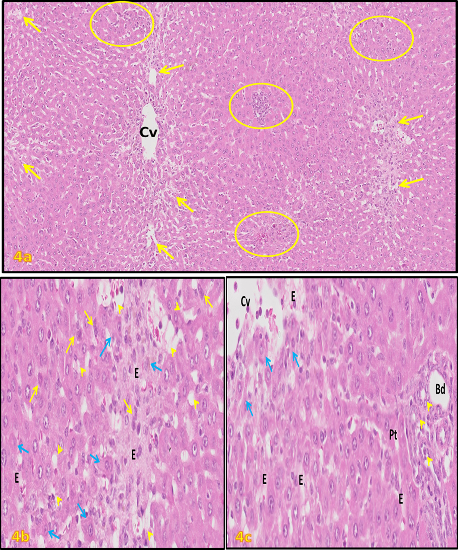
Figure (4a-c): L.M. of liver sections of adult rats treated with aqueous extract of kola nut with a dose of 10 mg/kg.BW/day for 60 days, then left the animals for normal recovery without treatment for 30 days (H. & E.). (a): destructed and lysis of hepatic strands (arrows) with strong inflammatory cell infiltrations (circles); H. & E. (100X). (b): High power from the above section showing vacuolization of hepatocytes (arrowhead), massive edema (E) accompanied by strand cell necrosis (blue arrows), pyknotic nuclei (yellow arrows); H. & E. (400X). (c): destructed central vein (Cv) through desquamation and necrosis hepatocytes (blue arrows), with edema (E); deformed and necrotic cells in portal tract (Pt) with proliferation (head arrows) of bile ducts (Bd); H. & E. (400X).
Table 1: The effect of daily injection of Kola nut extract (10 mg/kg, i.p.) and subsequent withdrawal on mean body weights (grams), mean liver weights (grams) and relative body weight (%) of treated versus control rats at the end of each experimental period.
Statistical analyses were performed between control (C=6) and treated (C=6) animals by using paired t-test. %: percentage of change form control; * = significant at P<0.05; ** = highly significant at P <0.001; Relative liver weight (%) = liver weigh (grams)/total body weight (gram) X 100. DISCUSSION In recent works, the liver structure of hepatocytes in the control rats is similar to what has been described in other mammals [7, 37, 40]. The histopathological findings in the current work indicated that treatment with aqueous extract of Cola nitida for 15 days led to severe congestion of the liver tissue, dilation of central veins, congestion with edema, invasion of inflammatory cells, and a significant decline in the Kupffer cells. These findings were consistent with previous studies, which determined the occurrence of cellular proliferation and the necrosis of Kupffer cells in some different cases of hepatotoxicity [41-43], or when treated with alcoholic extract of Kola nut [21]. Related studies also agree with our observations in mice liver that has been treated with a dose of 30 mg of caffeine extracted from the kola nut [44]. In addition, treatment with low doses of caffeine (5, 15, 30 mg/kg) promoted pneumonia and liver damage [45]. A study [46], confirmed that Cola nitida contains a high percentage of caffeine compared to its other components, which causes abnormal contraction of the smooth muscles in the blood vessels, which leads to congestion, erythrocytes stagnation and structure damage of blood vessels nourishing vital organs. The histological changes in treated rats for 60 days had an increased severity represented as vacuolar cytoplasmic degenerations, fatty infiltration, nuclear karyolysis, fibrosis of the portal tract, and bile duct proliferation. Corresponding records stated that treating rats with aqueous extract of Kola nut for 30 days caused cellular necrosis and cytoplasmic degenerations in seminiferous tubules [47]. The cytoplasmic vacuolations of brain and kidney tissues in treated mice have been attributed to the activity of secondary metabolites of Cola nitida extract [48, 49]. The obtained results are consistent with a study that reported a high dose (15 mg) of caffeine cause severe histopathological damage in renal tubules of rats appeared as glomerular inflammation, inflammatory invasion, cellular necrosis, and vascular degeneration [50]. In harmony with the study at hand, a significant increase (P <0.05) in the mean relative weight of the liver of rats treated with the Kola nut extract may be attributed to the portal and central veins congestion and fatty infiltration [44, 51]. In parallel with the results, it has been mentioned that impairment of fatty acid metabolism in liver cells leads to accumulation of neutrophilic lipid vacuoles in cells, which are not surrounded by membranes, and appear as faint empty vacuoles, and perhaps associated with hydropic degeneration [52]. However, the pathological changes in the nucleus indicate that the cells lose their functional efficiency [53]. Our result is in agreement with several prior researches, which indicated that the proliferation of bile ducts appears in the liver tissue associated with the process of cellular regeneration, inflammatory reactions, and fibrosis of connective tissues in the portal region [54, 55]. Research indicated that the cells lining the bile ducts are stem cells that proliferated when lysis or necrosis of hepatocytes occurred [56], which supports this study. Likewise, our results of the withdrawal group (G4) mentioned persistent histological damage as well as acute disturbance of the normal structure of liver tissue accompanied by a significant decrease (P <0.05) in the relative weight of the liver. These histological observations are supported with previous studies that reported prolong consumption of Kola nut reduces the concentration of superoxide dismutase and catalase and increases free radical generation, which enhances lipid peroxidation activity and oxidative stress results in the destruction of the cells, nuclear DNA, and can lead to some disease conditions or hepatotoxicity [57, 58]. In general, macrophages and lymphocytes are a sign of chronic inflammation, in which the macrophages ingest the pathogens and damaged tissue, while lymphocytes produce antitoxins and accelerate cellular healing [59]. Persistent inflammatory reactions after stopping the treatment in the present study attributed to the body's immune system reaction against the chemical damage, viral and parasitic infections, and agents harmful to cells and body tissues [52]. Several studies have discussed the formation of cytoplasmic vacuolations in mammalian hepatocytes indicated that the vacuoles represent early stages in the autolysis of cells after exposure to various pathogens and they prevent the accumulation of harmful substances and destructive elements in the cell and interfering with its main activities [7, 60, 61]. Whereas, the significant decrease (P <0.05) in the relative liver weight of rats in G4 was attributed to the severe degenerative changes and cellular necrosis caused by the Kola nuts treatment [44, 51]. CONCLUSION In conclusion, the present data and histopathological observations showed that long-term consumption of Cola nitida may lead to oxidative stress and result in hepatic tissue damage.
REFERENCES
|
|||||||||||||||||||||||||||||||||||||||||||||||||||||||||||||