International Journal of Pharmaceutical and Phytopharmacological Research
ISSN (Print): 2250-1029
ISSN (Online): 2249-6084

Centella asiatica L. is one of the plants that has been used since prehistoric times to cure several diseases. The main secondary metabolite in Centella asiatica L. which has the most role in various pharmacological effects is asiaticoside. Some of the following properties are healing skin diseases, wound healing, nerve weakness, brain cell stimulation, anticancer, antitumor, antioxidants, antibacterial, anti-function, anti-inflammatory, antituberculosis, allergy, antidepressants, and so on. Tissue culture is one method of botanical biotechnology that can produce plants with a character that can also increase the metabolism of secondary plants, including Centella asiatica L. Increased metabolism of secondary asiaticoside using the semi-quantitative PCR method. This method is based on genes that increase asiaticoside composition, as a proportion of the secondary metabolism. The gene that most influences asiaticoside formation is RNA UGT73AH1 (UDP-glycosilltransferase 73AH1). The purpose of this study was to analyze the expression of the UGT73AH1 gene in untreated and untreated Centella asiatica L. Analysis of gene expression was carried out based on the thickness of the band on the electropherogram which was implemented using the ImageJ application to produce AUC values. The results of the analysis using the semi-quantitative PCR method showed that Centella asiatica L. callus had the largest average AUC of 24,879.59, while plantlets were 23,780.04, and Centella asiatica L. plants were 7802.26, with ANOVA analysis produced a sig. value <0.05. This shows a significant average difference between groups of callus, plantlet, and conventional Centella asiatica L.
INTRODUCTION
Centella asiatica L. is one of the plants that have many benefits such as wound healing, brain cell stimulation, antioxidants, antibacterial, anti-function, anti-inflammatory, antituberculosis, alzheimer, antidepressant, etc. [1-6]. This plant has also been made into a variety of commercial products. Secondary metabolites in this plant that have a big role in pharmacological effects are the centellosides, one of which is the asiaticoside compound [6]. The many pharmacological effects of these compounds make an increase in the needs of asiaticoside to meet the availability of asiaticoside as a source of treatment materials for various diseases, both in commercial and non-commercial forms [7-9].
Research that has been done before, shows that the application of fertilizer and extraction of ordinary plants produces less maximal asiaticoside, so we need another method to get higher asiaticoside. Besides, the propagation of secondary metabolites by conventional methods also has the possibility of differences in the quality and quantity of the contents of plants because they grow in different places, nutrients, light, and other conditions [10].
Problems that exist in conventional methods make the need for another method that can produce plants with a uniform quality but has a high content, namely tissue culture. Plant tissue culture can increase the content of secondary metabolites with minimal space and time requirements [11-13]. Although this method requires conditions that are always sterile, it has many advantages, namely a high rate of multiplication, the growing environment can be controlled as needed, plants are not affected by regional or seasonal variations, and can produce plants with better genetic engineering [14, 15].
The increase of asiaticoside in tissue culture can be analyzed using the semi-quantitative PCR (Polymerase Chain Reaction) method [16]. This semi-quantitative analysis is based on the expression of genes that play a role in the synthesis of secondary metabolites. The expression of these genes can be detected by isolating the mRNA that most influences the biosynthesis of the secondary metabolite. The most influential gene in the biosynthesis of asiaticoside is UGT73AH1 (UDP-glycosilltransferase 73AH1) RNA [17, 18]. Analysis using PCR has several advantages, namely the required small sample size, specificity to certain species, high sensitivity, the potential to confirm the identification of botanical material in the intact or truncated form [19-22]. This is the researcher's goal to be able to increase the production of asiaticoside in Centella asiatica L. through tissue culture techniques, so that it is possible to meet the needs of medicinal raw materials for various diseases, with effective quantification methods.
MATERIALS AND METHODS
Centella asiatica L. tissue culture
The media composition for plantlet and callus culture was Murashige and Skoog (MS) media 4.43 g / L, agar 7 g / L, sugar 30 g / L, and aquadest. The difference in composition for the plantlet and callus media is in the growth hormone, whereas for the growth hormone of plantlets using the BA 0.5 ml / L while for callus using the BAP hormone 1 mg / L, the NAA hormone 1.5 mg / L. Media pH is 5.8-6.0, adjusted using 1 N HCl or 1 N NaOH. Then the media that has been mixed boil and then poured into a bottle with ± 1 cm left, close the bottle, sterilized using an autoclave, then compacted. All stages are carried out in a sterile and aseptic manner [11, 23].
The explant used for the manufacture of callus were leaves from plantlet (obtained from Esha Flora, Bogor, Indonesia), while the leaves of Centella asiatica L. were used as a comparison of asiaticoside levels from Centella asiatica L. tissue culture. Explant injured first then placed on a media that is sterile and solid. Incubation was carried out at a temperature of 26 ° C ± 2 ° C and humidity of 55-60%. Incubation for callus forms was carried out for 2 months. The result of this incubation will be in the form of a callus which will then be isolated from UGT73AH1 mRNA [11, 24].
Detection of mRNA UGT73AH1 Gene
RNA isolation of Centella asiatica L. as well as the culture results of plant tissue using the Technical Manual SV Total RNA Isolation System (Promega) procedure. The principle of this isolation is cell wall lysis, pre-precipitation, and purification. The main stages in this process are lysis by grinding after the addition of liquid nitrogen and the addition of RNA lysis buffer and dilution buffer, then pre-precipitation is done by the spin (centrifugation) method, and purification is done by adding DNAse stop solution and RNA wash solution. The result of this process is a liquid containing an RNA template as one of the components that will be used in the RT-PCR process.
RNA can be amplified if the RNA is converted to cDNA (complementary DNA). The change to cDNA will be carried out with the enzymatic process Reverse Transcriptase (RT). This process uses the Access QuickTM Master Mix (Promega) kit, with the addition of forwarding primer components, reverse primer, RNA templates, nuclease-free water, and AMV reverse transcriptase. The PCR conditions used were initial denaturation: 45 ° C 45 minutes, followed by 40 cycles of amplification for 15 s at 95°C, for 15 s at 58°C, and 30 s at 72°C, post PCR 4°C.
Primer design is done by searching the literature, where the primer sequence is obtained from the journal [17] in the order of 5 'to 3' forward, are GCATCATGTCTGAAGATGAGG and sequence 5 'to 3' reverse which is GCTTAATGCTAACCTATCCTT. This primer sequence is evaluated using software both online and offline (Snapgene). The primer parameters evaluated are Tm (melting temperature), % GC, nucleotide length, and the presence of dimer primers. Next, the primary projection of Centella asiatica L. was also used by the Nucleotide BLAST software at the NCBI website.
Characterization of RT-PCR (cDNA) amplicons was carried out using gel base electrophoresis with the addition of 0.5 μl diamond for every 40 ml of agarose. A 5 μL sample and 2 μl loading dye were pipetted and homogenized, then put into a well. 2 μL ladder of 100 bp is added to one of the wells. The system is installed on the 100-volt range with a duration of 30 minutes with the sample running from the negative pole to the positive pole. Judging by the results above the Blue light Mupid using a UV filter. The results of this process are in the form of electrophoregram which will then be quantified using the ImageJ application.
Semi-quantitative using imagej
The electropherogram is quantified using the ImageJ application, where the thickness of the tape on the electrophoregram photo is a projection of the AUC (Area Under Curve) value. The data is a semi-quantitative result of the nucleotide levels that have been copied by PCR, which is a picture of the asiaticoside levels of the sample [25, 26].
RESULTS AND DISCUSSION
Centella asiatica L. tissue culture
The media content greatly influences the results in tissue culture, especially color and shape [15]. The composition of MS media (murashige skoog), agar, sucrose, and aquadest were given with the same final volume, both on the callus and planlet media. But the difference in composition lies in the addition of growth hormone (Table 1).
Table 1. Callus and Planlet Media Composition
|
Ingredient |
Callus (/L) |
Planlet (/L) |
|
Media MS |
4.43 gram |
4.43 gram |
|
Agar |
7 gram |
7 gram |
|
Sucrose |
30 gram |
30 gram |
|
Growth Hormone |
BAP: 1 mg NAA: 1.5 mg |
BAP: 0.5 mg |
|
Aquadest ad |
1 L |
1 L |
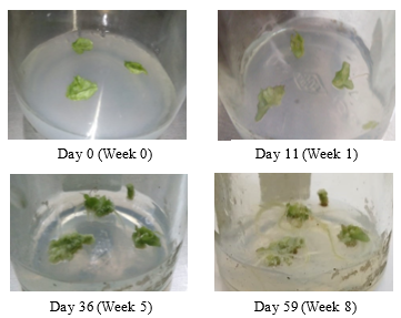
The presence of auxin in tissue culture media influences the process of cell division, cell elongation, apical dominance, adventitious root formation, somatic embryogenesis, increasing rhizogenesis, and callus formation. Whereas the cytokine hormone influences cell division, stimulates the initiation and development of buds in vitro, inhibits root formation, and induces adventitious bud formation. So that the presence of the hormone cytokines in plantlet growth media, will induce the formation of plants as a whole compared to the media in callus growth [15, 27]. Callus growth can be seen in Figure 1.
Callus growth will be followed by callus discoloration [11, 28]. As the callus grows, it starts from light green (week 1), dark green with little white tissue (week 5), and green with a little brown tissue (week 8). The change in color of the callus can be influenced by light, the presence of pigmentation, and plant parts were taken to be used as explants [29]. Explants that are green but produce callus colors that are white or brown indicate chlorophyll degradation [30]. Whereas, if the explants that are green produce a green callus color, it indicates that the presence of chlorophyll in the callus tissue. The green color can also be caused due to an increase in the cytokine hormone because cytokinin can inhibit the destruction of chlorophyll and activate the metabolic process and synthesis. While the brown color of the callus can be caused by increasing callus age, the longer is the callus's age, the darker is the brown [31, 32].
|
|
|
Figure 1. Callus Growth for 8 Weeks |
Callus texture can be observed to determine whether the callus is still actively dividing or not. In general, based on the texture of the callus can be divided into 3 types, namely, compact texture (non-friable), crumb texture (friable), and intermediate texture (a combination of crumb and compact texture). Callus with a compact texture has a strong collection of cells, both on the inside or the surface. Callus with crumb texture has a collection of cells that are easily separated either on the inside or the surface of the callus. While the callus with an intermediate texture has a strong collection of cells on the inside, but crumbs on the outside or surface [31].
In this study, the texture of the callus grew more compact (Figure 1). This can be caused by hormones or growth regulators added to the callus growth media. Where the callus given the cytokine hormone (in this study used BAP) will produce a compact callus texture. In addition, the auxin hormone together with the cytokine hormone can also affect the potential of water in cells, where the absorption of water that carries hormones or nutrients from the growing media into cells will increase so that the callus becomes stiffer [33].
Based on Loc (2013) research on the asiaticoside content in the callus of Centella asiatica L., the compact texture had a higher secondary metabolite content compared to the crumb textured callus [34]. This can be caused because the callus with a compact texture has a solid cytoplasm and a large cell nucleus [35].
Detection of UGT73AH1 mRNA Gene
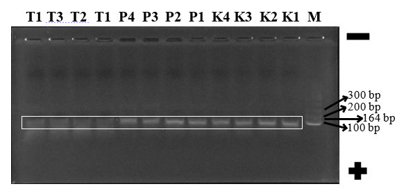
In the results of the primer analysis, the product size of the amplification was 164 bp, so the asiaticoside band location was at the length of the base pair. The UGT73AH1 gene is parallel to the marker band whose position is between 100 bp and 200 bp (Figure 2).
|
|
|
Figure 2. Electrophoregram Amplicon UGT73AH1 Gene in Centella asiatica L. |
Information:
|
M: Marker (ladder 100 bp) K1: Callus 1.1 K2: Callus 1.2 K3: Callus 2.1 K4: Callus 2.2 |
P1: Planlet 1.1 P2: Planlet 1.2 P3: Planlet 2.1 P4: Planlet 2.2 |
T1: Plant 1.1 T2: Plant 1.2 T3: Plant 2.1 T4: Plant 2.2 |
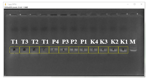
The results of electrophoresis in the form of electrophoregram were then analyzed using the Image J application (Figure 3), where each band will produce AUC (Area Under Curve) based on the thickness of the tape. The thicker the band, the higher the AUC will be. High AUC correlates with the level of secondary metabolites of plants tested, in this case, the level of asiaticoside from the UGT73AH1 Centella asiatica L. gene. The semi-quantitative analysis of AUC can be seen in Table 2.
|
|
|
Figure 3. Semi-Quantitative Analysis Using the Image J Application |
Table 2. Asiaticoside AUC (Area Under Curve) Value-Based on Electrophoregram UGT73AH1 Centella asiatica L. Gene Expression Results
|
No. |
Sample |
AUC |
No. |
Sample |
AUC |
|
K1 |
Callus 1.1 |
26.919,354 |
P3 |
Planlet 2.1 |
23.675,655 |
|
K2 |
Callus 1.2 |
25.381,040 |
P4 |
Planlet 2.2 |
24.893,333 |
|
K3 |
Callus 2.1 |
21.891,676 |
T1 |
Plant 1.1 |
8970,877 |
|
K4 |
Callus 2.2 |
25.326,304 |
T2 |
Plant 1.2 |
5387,777 |
|
P1 |
Planlet 1.1 |
23.482,454 |
T3 |
Plant 2.1 |
6356,513 |
|
P2 |
Planlet 1.2 |
23.068,697 |
T4 |
Plant 2.2 |
10.493,889 |
The results of this study can be seen as an increase in callus and planlet AUC, from the expression of the UGT73AH1 gene, which is responsible for the formation of secondary metabolites of asiaticoside in Centella asiatica L., where AUC is correlated with asiaticoside levels.
Increased levels of secondary metabolites in tissue culture products are strongly influenced by growth hormone. Growth hormone regulates various physiological and morphological processes in plants and is also known as a plant growth regulator or phytohormone. The content of individual auxin such as NAA and cytokines like BAP in the media used influences the growth and regulation of cell metabolism. Cytokinins are known to increase the production of secondary metabolites and play an important role in cell differentiation and subcellular differentiation [15, 36, 37].
In addition to growth regulators, plant tissue culture in this study, and in general using MS media (Murrashige and Skoog) containing sucrose, where sucrose is a carbon source that has a significant impact on metabolite yields from in vitro culture. MS media also contain nitrogen, where nitrogen is an important component of several culture media [15, 38].
This study also showed that on average, callus produced asiaticoside 1.46 times greater than plantlets and 3.19 times greater than Centella asiatica L. plants without any treatment. This can be caused because the source of explants for callus growth used is from selected plant parts that contain the most asiaticoside compared to other parts, namely leaves. So that the cell growth that grows in the callus is only a cell from the leaf. While plantlets, although sourced from leaves, in the end, the asiaticoside content will spread to other plant parts besides the leaves. Also, the structure of callus is no more complex than ordinary wild plants and plantlets, and the callus also has a smaller area and volume than ordinary plants and plantlets, so that growth regulators added will be more easily absorbed and then spread to all parts of the callus.
CONCLUSION
Tissue culture in both callus and plantlet forms can increase UGT73AH1 gene expression in Centella asiatica L. and increase the content of secondary metabolites of asiaticoside compared to ordinary plants.
Acknowledgments: We would like to thank Mrs. Rosydiati Arasyd for helping with the tissue culture.
Conflict of interest: None
Financial support: Thanks to LPPM Universitas Bhakti Kencana for providing research funding.
Ethics statement: None.
.