International Journal of Pharmaceutical and Phytopharmacological Research
ISSN (Print): 2250-1029
ISSN (Online): 2249-6084
|
Study of the Ossification Centers of Thorax in Partridge “Alectoris Chukar” After Hatching by Radiographic and Staining Techniques
Masoud Sahimirad1, Hassan Gilanpour1, Mohammadreza Paryani2*, Abbas Veshkini3, Bijan Radmehr4 |
|
1 Department of Basic Sciences and Hygiene, Science and Research Branch, Islamic Azad University, Tehran, Iran. 2Department of Basic Sciences, Faculty of Veterinary Medicine, Karaj Branch, Islamic Azad University, Karaj, Iran. 3Department of Clinical Sciences, Science and Research Branch, Islamic Azad University, Tehran, Iran. 4Department of Basic Sciences, Faculty of Veterinary Medicine, University of Tehran, Tehran, Iran. |
ABSTRACT
The purpose of this study is to consider the thoracic cavity ossification centers, time and formation and completion of bones. In this paper, 50 fertilized eggs of Partridge were placed inside the incubation unit for hatching. Spilling of them at 2 birds for radiography and modified double staining Alizarin Red and Alican Blue, in the first week of two birds (first and seventh days) and then at the end of the second week, third, fourth, fifth, sixth, seventh, eighth, ninth, respectively. The results of this study showed that the ossification centers in some of the bones such as thoracic vertebrae, ribs, and craniolateral process and caudolateral process of the sternum are apparent in the embryonic stage, but the ossification centers in the body of the sternum and sternal crest will be ossified after hatching up to ninth week. It may be said that all of the thoracic areas are ossified and skeletal development is completed on day 70 after hatching.
Key Words: Ossificationcenters, Thorax, Hatching, Staining, Partridge.
The studies on the skeleton system of the embryo and adult bird in the anatomy level, embryology, histology are important from different aspects. Partridge meat with unique nutritional capabilities than to other animal proteins and hen, including higher protein and lower cholesterol and early digestion, is a good source of an animal protein supply, especially in older adults and children.
Evolutionary skeletal growth in birds has always been a topic of interest.
Many studies have been conducted on the chicken, quail, turkey, a duck, with specific aspects of the development of the Pelvic limb skeleton, vertebral column, stifle joint [1, 2] or growth of a particular bone, for example, “tibia - tarsus ” or “tarsometatarsus”. [3, 4] The Chukar Partridge race (Alectoris Chukar) was a popular species of Partridge, which is used for breeding. The weight of the bird is 600g, and it lays 40-50 eggs in the breeding season.
Reviews show that the study on different parts of Partridgesuch as of the wing, [5] leg skeleton, [6] spleens, [7] thymi, [7] liver, [8] esophagi, [9] stiflejoint [10] has been done by different researchers in terms of the histology and anatomy. This study is very important since the practice of breathing in birds and hunter birds especially that have a high metabolism and they spend a lot of energy flying, and on the other hand, the thoracic cavity also plays an important role in the respiratory function. Therefore, the purpose of this study was to evaluate the time and extent of bone formation in the thoracic cavity ossification centers and their completion.
MATERIALS AND METHODS:
In this study, 20 Chukar Partridge chicks with unknown sex were used for radiographic and staining examinations. For this purpose, 50 Partridge fertilized eggs that were produced from the Partridge breeding farm are incubated in the incubator according to its instruction (Twin cocks machine, cocks company). The eggs were kept in the apparatus at temperature (37) and humidity (55-56%). Twenty chickens were selected for the study, taking into account the fertilized eggs and dead embryos and chicks.
Chicks were kept in the same condition, in terms of food rations, temperature, and humidity. The diet consisted of starter food. For this study, two birds of them were randomly sampled on the first day (after hatching) at the end of the first weeks, second, third, fourth, fifth, sixth, seventh, eighth, ninth, respectively. Type of device was digital radiography unit(mAs 0.6, KVp 50, FFD 75 cm, Samsung Flat Panel – Vanak imaging center). Right lateral and ventrodorsal radiographs were taken. Samples were stained using an Alizarin Red and Alcian Blue staining technique [11] and different parts of the thoracic cavity including thoracic vertebrae, ribs and sternum were examined by a stereomicroscope (model ZMZ1, the company of cocks machine) and the specimens were photographed. The study design and ethical approval were obtained by the Faculty of Veterinary Medicine, Science and Research Branch, Islamic Azad University, Tehran, Iran (IR.IAU.SRB.REC.1396.153).
RESULTS
The resulting data from the study of different parts the thoracic cavity of Partridge showed (Table1, 2). On the first day, only ribs and vertebrae were ossified completely (Figure 1a) and examination of the stained specimens revealed the body and arch of vertebrae were ossified and the only spinous process is remained as cartilaginous (Figure 3a). The thoracic vertebrae have 5 ossified ribs. The part of vertebral and sternal ribs. Both ends of the sternal rib have remained as cartilaginous. The uncinate process of rib 1, 2, 3 long remains cartilaginous at the two ends and in the middle of the bone. The sternal part of the fifth rib is cartilaginous and is connected to the sternal rib of the fourth rib which is considered to be a false rib. The sternum is completely cartilaginous and the craniolateral process of the sternum is ossified and the only proximal end has remained cartilaginous.
The manubrium is transparent and goes through the process of ossification. The caudolateral processes ossified but the proximal end is cartilaginous (Figure 3b).
Table 1. The Time of observing of Thorax Bones and its various structures in the radiography.
|
Bone |
D1 |
W1 |
W2 |
W3 |
W4 |
W5 |
W6 |
W7 |
W8 |
W9 |
|
Thoracic vertebrae |
+ |
+ |
+ |
+ |
+ |
+ |
+ |
+ |
+ |
+ |
|
Notarium |
- |
- |
- |
- |
- |
- |
- |
- |
- |
+ |
|
Vertebral rib |
+ |
+ |
+ |
+ |
+ |
+ |
+ |
+ |
+ |
+ |
|
Sternal rib |
- |
-+ |
-+ |
+ |
+ |
+ |
+ |
+ |
+ |
+ |
|
Uncinate process |
- |
- |
- |
+ |
+ |
+ |
+ |
+ |
+ |
+ |
|
Body of sternum |
- |
- |
+- |
+ |
++ |
+++ |
++++ |
+++++ |
++++++ |
+++++++ |
|
Caudo-lateral process of sternum (Medial Pro.) |
- |
+ |
+ |
+ |
+ |
+ |
+ |
+ |
+ |
+ |
|
Caudo-lateral process of sternum (Lateral Pro.) |
- |
+ |
+ |
+ |
+ |
+ |
+ |
+ |
+ |
+ |
|
Cranio-lateral process of sternum |
- |
+ |
+ |
+ |
+ |
+ |
+ |
+ |
+ |
+ |
|
Keel |
- |
- |
- |
+ |
++ |
+++ |
++++ |
+++++ |
++++++ |
+++++++ |
The number of marks (+) represents the process of orthogenesis.
Table 2. The Time of observing of Thorax Bones and its various structures in the Staining.
|
Bone |
D1 |
W1 |
W2 |
W3 |
W4 |
W5 |
W6 |
W7 |
W8 |
W9 |
|
Thoracic vertebrae |
+ |
+ |
+ |
+ |
+ |
+ |
+ |
+ |
+ |
+ |
|
Notarium |
- |
- |
- |
- |
- |
- |
-+ |
-+ |
-+ |
+ |
|
Vertebral rib |
+ |
+ |
+ |
+ |
+ |
+ |
+ |
+ |
+ |
+ |
|
Sternal rib |
+ |
+ |
+ |
+ |
+ |
+ |
+ |
+ |
+ |
+ |
|
Uncinate process |
+- |
+- |
+- |
+ |
+ |
+ |
+ |
+ |
+ |
+ |
|
Body of sternum |
- |
+- |
+- |
+ |
+ |
+ |
+ |
+ |
+ |
+ |
|
Caudo-lateral Process of Sternum (Medial Pro.) |
+- |
+ |
+ |
+ |
+ |
+ |
+ |
+ |
+ |
+ |
|
Caudo-lateral process of sternum (Lateral Pro.) |
+- |
+ |
+ |
+ |
+ |
+ |
+ |
+ |
+ |
+ |
|
Cranio-lateral process of Sternum |
+ |
+ |
+ |
+ |
+ |
+ |
+ |
+ |
+ |
+ |
|
Keel |
- |
- |
+- |
+ |
+ |
+ |
+ |
+ |
+ |
+ |
In the study of radiography of the first-week chick, the ossification process is progressive, bone density has increased and the cervical and thoracic vertebrae are visible in the ventrodorsal view (Figure 1b). In the lateral view, the medial and lateral part of the caudolateral process is quite clear (Figure 1c). In the stained specimens, it was found that the head of ribs is ossified. The thoracic vertebrae are not fused. The vertebral and sternal sections of the ribs are ossified except for the last two thoracic ribs. Uncinate process of the last cervical rib and the three thoracic ribs are predominantly bone and the fourth cartilaginous and the last two ribs have no uncinate process (Figure 3c).
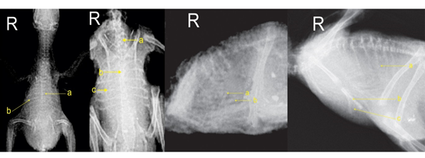
Fgure 1: 1A) Dorsal view at day 1 (a) Thoracicvertebrae (b) Rib, 1B) Ventro - dorsal view at week 1. (a) Cervical Vertebrae (b) Thoracicvertebrae (c) Rib, 1C) Lateral view at week 1. (a) Lateral process of the caudolateral process (b) Medial process of the caudolateral process
The middle part of the sternum is ossified. The craniolateral is also ossified. The medial and lateral part of the caudolateral process are ossified and only the proximal end is cartilaginous and is also visible (Figure 3d). Radiography of chick has been similar to one another at the end of the second and third weeks. The middle portion body of the sternum is ossified. Sternal crest goes through the process of ossification (Figure 1d). The keel is ossifying progressively. The stained specimens it was revealed that the more advanced bone process, the thoracic vertebrae closer together, and the ventral crest of the cervical vertebrae and thoracic vertebrae were seen cartilaginous. The vertebral and sternal ribs are ossified and the process of ossification internal ribs is increasing from cranial to caudal. The middle part of the sternum is ossified and sternal crest goes through the process of ossification (Figure 3e).
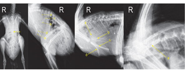
Fgure 2: 1D) Lateral view at week 3. (a) Rib (b) Body of sternum (c) Keel, 2E) Ventrolateral view at week 4. (a)Thoracic vertebrae, 2F) Lateral view at week 4. (a)Vertebral rib (b)Sternal rib (c) Uncinate process (d) Body of sternum (e) Keel
2G) Lateral view in week 8. (a) Craniolateral process (b) Caudo - lateral process (c) Body of sternum (d) Keel, 2H) Lateral view at week 9. (a) Notarium
In the radiographs of the end of the fourth-week chick, the thoracic vertebrae are fully defined, and the bone density is increased from cranial to caudal, but it has not been fused yet (Figure 1e). The vertebral and sternal ribs are ossified and the uncinate process is distinct and ossified has seen completed. The progress of bone in the sternum increasing and the first part of sternal crest is ossified (Figure 1f). In the stained specimens, the cranial and middle portions of the sternum body except for the cranial border bone, and where the craniolateral and caudolateral processes are connected to the sternum and each other are seen cartilaginous (Figure 3f). The cranial part of the sternal crest is ossified and the craniolateral process is transparent and goes through the process of the ossifying process(Figure 3g).
The radiographs of chicks at the end of the fifth and sixth weeks were similar to the chick of the fourth week and the ossifying process and density of bone are also increasing. In the stained specimens of the fifth-week attachment of craniolateral and caudolateral processes to the sternum and each other are ossified (Figure 3h). In chicks at the end of the sixth week, the ventral crest of the last cervical vertebrae and thoracic vertebrae which were cartilaginous began to become ossified. Part of the manubrium is seen ossified (4i). In radiographs of the chick at the of the seventh week is completely defined, thoracic vertebrae, vertebral and sternal ribs. The process of the density of the sternum and sternal crest ossification is also progressive. The craniolateral and caudolateral processes are completely ossified and defined (Figure 2g). In the stained specimens, it was observed that the cranial border of the sternum and craniomedial process of the sternum goes through the process of ossification (Figure 3j). The radiographs of chicks at the eighth week are according to the seventh week, these changes are progressive, and is added to the ossified density of different sections. In the stained specimens, the spinous process of the cervical vertebrae and thoracic vertebrae is ossified but has not been fused and they are seen as cartilaginous (Figure 3k).
In radiographic of the chick at the ninth week has been fusing the cervical vertebrae and thoracic vertebra so the aquarium is formed (Figure 2h). In the stained specimens is also observed that bone density is progressive in the different part of the thoracic cavity. The spinous process of the last cervical vertebrae and the first three thoracic vertebrae are fused, but the proximal end of the spines also as cartilaginous (Figure 4m). The cranial border of the sternum goes through the process of ossification (Figure 3n).
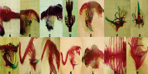
Fgure 3: 3A) Dorsal view at day 1. (a) Spinous process
3B) Lateral view at day 1. (a) Uncinateprocess (b) 5th Rib (c) Body of the sternum (d) Craniolateral process (e) Cranio - medial process (f) Caudolateral process
3C) Lateral view in week 1. (a) Head of the rib (b) Thoracic vertebrae (c) Uncinate process
3D) Lateral view in week 1. (a) Body of the sternum (b) Cranio - lateral process (c) Caudolateral process
3E) Lateral view at week 3. (a) Thoracic vertebrae (b) Ventral crest (c) Body of sternum (d) Keel
3F) Lateral view at week 4. (a) Cranial border of the sternum (b)Attachment site of Cranio - lateral and Caudo - lateral processes to the sternum and each other
3G) Ventral view at week 4. (a) Keel (b) Craniomedial process
4H) Lateral view in week 5. (a) The attachment site of Cranio - lateral and Caudo - lateral processes to the sternum and each other
4I) Lateral view in week 6. (a)Ventral crest (b) Craniomedial process
4J) Lateral view in week 7. (a) Cranial border of the sternum (b) Craniomedial process
4K) Lateral view in week 8. (a)Spinous process
4L) Dorsal view in week 8. (a)Spinous process
4M) Dorsal view in week 9. (a)Thoracic vertebrae (b) Spinousprocess
4N) Lateral view in week 9. (a) Cranial border of the sternum
DISCUSSION:
Skeletal growth in birds has always been a topic of interest. The first studies about bone histogenesis were performed by Brachet in the year (1893), [12] but done in more comprehensive form by Fell in the year (1925), [3] and the studies were followed by Hegested and Wolbach in the post-hatching period. [13]
Despite the importance that the thoracic cavity inbreathing and the flying bird which is high in metabolism and is used to carry out a lot of energy of fly, the study is relatively rare concerning the ossifying and the process of skeletal ossification and thoracic cavity bones.
In various studies ranging from ossification centers at the time of embryonic evolution, the best study was carried out by Hamilton (1952) and Romanoff (1960) that it has been done on the hen at all stages of mesenchymal, cartilaginous and ossification. [14, 15]
In 2016, a study was conducted on the ossifying trend in the hybrid hen before and after hatching by Bambang Retnoaji et al. [16] In this study, 30 eggs were used and embryos were evaluated from day 5 of incubation until the seventh week after hatching. This study showed that osteogenesis begins on day 10 of incubation and that ossification centers appear earlier in vertebrates and ribs compared to partridge embryos.
In 2019, a study was conducted on the ossification process of vertebrae; thoracic limbs and sternum before and after hatching in the Japanese quail by Pourlis et al. [17] The study revealed that ossifying was observed in the thoracic vertebrae at 10 and 11 incubation day trough ribs began to become ossified at day 7 of incubation. The uncinate process began to become ossified on day 15. Craniolateral and caudolateral processes indicate the ossification at day 13 and 14 of incubations, respectively. The body of the sternum and sternal crest began also to become ossified after the hatching. It was observed in the present study that the sternum began to become ossified at the end of the first week and the sternal crest at the end of the second week.
From the study of the above studies, we have concluded that the study fulfilled were only at the embryonic period and the best study was performed at the time of hatching and then by Hogg (1980), which was also on the hen [18]. The results of the study of Hogg shows that the vertebrae have two ossification centers, one of the vertebral arch and the other in the vertebral body.
In the ribs, two ossification centers appeared separately in the vertebral and sternal sections of the ribs. The ossification centers are apparent in uncinate processes at the time of hatching. There are 5 ossification centers in the sternum, one in the body and two in the craniolateral and caudolateral processes on each side.
In the present study, the first ossification center was observed in the sternum and sternal crest body during the third week, which was observed in the radiographic examination of the sternal body and sternal crest. At the end of the third week of ossification in the processes of craniolateral and caudolateral were also observed the osseous on the first day, but in radiography, at the end of the second week, they were found ossified.
In 2007, studies were conducted on the ossification of the sternum and long bones of pelvic limbs skeleton in the rooster and hen by Breugelmans et al. [19] In this study, about 20% of the sternum body and sternal crest remained cartilaginous till 14 weeks in the rooster and 20 weeks in the hen and the process of ossification happened slowly. The findings of this study are consistent with the present study that continues until the ninth week the process of the ossification process in the body of the sternum and sternal crest.
In 2019, a study was conducted on the pectoral girdle ossification centers in the birds by Dadashpoor Mohammad et al. [20] In that study, radiographs of sternum were not observed in any of the birds up to day 7, but it was observed in specimens from day 14 to later. In the present study at day 7 body of the sternum of Chukar Partridge was found fully ossified in the stain specimens. However, in the previous studies, there is no investigation on ossification centers before and after hatching. In this study, ossification centers have been studied during and after the hatching by using two radiographic technique and staining and also a comparison has been done between the thoracic cavity skeleton in ChukarPartridgerace and hen. In the studies conducted on the Partridge chicks’ from hatching day until the end of the ninth week using both staining and radiographic technique, the result was that the thoracic cavity including thoracic vertebrae, ribs, and sternum, would travel progressively week by week. In radiographs generally study the bone density, the presence or absence of a structure, but in stained specimens, the different parts of the specimen are examined in greater detail.
In the study of the radiographs that had been made from the first day chicks, the thoracic vertebrae were well visible in the stained specimens, as well as the body parts of the vertebrae are quit ossified, but the process of the spine has remained cartilaginous. The vertebral portion of the ribs is also cartilaginous, but the distal end of the vertebral ribs and the proximal end of the sternal ribs are still cartilaginous. The distal end of the sternal ribs is still cartilaginous in the joint with the sternum. The uncinate processes of ribs were not observed in the radiographs whereas in the ribs stained specimens of 1,2,3 had a relatively long uncinate process that was found in both ends cartilaginous and in the middle section as a bone. In radiographs, the body of the sternum and the sternal crest was a completely cartilaginous structure that was found in the stained specimens, as well. In radiographs, craniolateral and caudolateral processes are seen with low density whereas in the stained specimens they are seen as osseous, and only the ends of each process are cartilaginous. It was not observable the craniolateral process, whereas in the stained specimens was observed transparent and it goes through the process of ossification. In radiographs, the chick at the end of the first week's uncinate processes are not yet visible, but they are highly visible in the stained specimens. It is observed in the radiographs of the sternum, whereas in the stained specimens it is observed that the cranial part of the sternum is ossified. At the end of the second week, the uncinate processes of ribs are not seen while in the stained specimens of bone. In radiographs, sternal crest with low density is seen that showing the process of ossification whereas in the stained specimens cranial part of sternal crest is seen bone. At the chick of the end, the third-week uncinated process was observed with low density, whereas in the stained specimens they are perfectly osseous, and only a very small part of ends of it remained cartilaginous. In radiographs, the attachment site of the craniolateral and caudolateral are not visible processes to each other and the body of the sternum. In the stained specimens, these parts have remained cartilaginous. In radiographs, the craniolateral process of the sternum was not observed whereas in the stained specimens transparently was observed and it goes through the process of ossification. At the chick of end the fifth week attachment site of the craniolateral and caudolateral processare not explicitly visible to each other and the sternum body is clearly, whereas in the stained specimens is seen as an osseous. At the chickof end the sixth week ventral crest of the last cervical vertebrae and thoracic vertebrae is not clearly visible, whereas in the stain showing the process of ossification. At the chick of end the seventh week and eighth week there will also be progressive changes that increase the bone densitywhereas in the stained specimens the body of the cervical vertebrae and thoracic vertebrae were fused. But in the spinous process is separated from one another. In the ninth week radiographs, it was observed that the last cervical vertebra was attached to the first three vertebrae and formed a notarium structure, which was also observed in the stained specimens.
Finally, it can be concluded that the lack of structural observation in radiography does not indicate that it is not a structure. It was a cartilaginous structure or it goes through the process of ossification, that may not be observed, and can be found in radiographs by ossifying and increase of the bone opacity and bone densitometry. Therefore, in this study, it is used modified double staining Alcian Blue and Alizarin red until the ossification center examinations are done carefully.
Although variation occurs at different times for the bone density, the primary cartilaginous and the osteogenesis in the thoracic cavity it is seen that evolutionary growth of the thoracic skeleton in Partridge is very similar to the quail and the chicken.
Comparison of the process of ossification in Partridge with a quail and a hen, it is clear that skeleton elements in each of the three birds are similar together, although evolutionary growth in Partridge is longer than that of chickens and longer than that of quails, the final form of the thoracic cavity is similar in all three birds.
Therefore, according to the growth of the thoracic cavity of Partridge, the chicken and quail seem to be classified properly in the Galiform class [21].
ACKNOWLEDGMENTS:
We appreciate very much for Dr. Hossein Eric Aghaji and Dr. Azadeh Zakeri for the great of their contribution to this paper.
Conflict of interest:
No conflicting interests and no funding in connection with this paper are applicable.
REFERENCES


