International Journal of Pharmaceutical and Phytopharmacological Research
ISSN (Print): 2250-1029
ISSN (Online): 2249-6084

|
Sacrum Ultrasonography: A Viable Alternative to Radiography in Infants with normal Sacrum? Maryam Riahinejad1, Mohsen Masoumi2*, Maryam Farghadani3, Mehrdadreza Hosseinpoor4, Parvaneh Changanian5 |
|
1 Assistant professor, Department of radiology, medical school of Isfahan University of Medical Sciences, Isfahan, Iran, 2Resident of Radiology, Department of radiology, medical school of Isfahan University of Medical Sciences, Isfahan, Iran, 3 Associated professor. Department of radiology, medical school of Isfahan University of Medical Sciences, Isfahan, Iran, 4 Aassociated professor, department of pediatric, medical school of Isfahan University of Medical Sciences, Isfahan, Iran, 5 Department of radiology, medical school of Isfahan University of Medical Sciences, Isfahan, Iran. |
ABSTRACT
Aim: Radiography, as the main method for diagnosis of abnormalities in sacrum, exposes infants to ionizing radiation. This study was aimed to compare the agreement of sacrum ultrasonography with radiography for the diagnosis of sacrum abnormalities. Methods: 129 infants under the age of six months, who were candidate for abdominal and/or pelvis radiography were assessed. Sacrum ultrasonography and radiography were performed by a single radiologist using the same devices. The sacral ratio was calculated for all patients based on ultrasonography and radiography findings and agreement between the two methods was calculated by Bland Altman's chart. Results: The mean of sacral ratio in studied infants based on radiography was 0.70±0.11 and based on ultrasonography was 0.72±0.05. Based on Bland Altman the mean difference between ultrasonography and radiology was 4.6 cm (confidence intervals of 8.18 ± 5.6). Conclusion: Sacrum ultrasonography could be safely used in the investigation of sacral ratio to detect sacrum abnormalities in infants, although, additional assessment are required for accurate assessment of ultrasonography with radiography.
Key Words: Sacrum, Sacral ratio; Sacrum ultrasonography; Sacrum radiography.
INTRODUCTION
Sacrum is a large triangular bone that is composed of five sacral vertebrae in neonates and gradually ossify and fuse to form the adult sacrum in the third decade of life [1]. It supports pelvic bones and maintains pelvic stability and provides the necessary conditions for conveying body weight to the pelvis and also produces significant movement in the pelvic during fertility [2]. The upper part of the sacrum is bulk because it transfers body weight to the pelvis, and the lower part does not receive much weight, and it is cone-shaped and narrow [3].
The sacral ratio, which is calculated by comparing sacrum size with fixed bony parameters of pelvis, is known as a main index to assess the sacrum abnormalities. This index was proposed in 1995 as a reliable tool to evaluate sacral development in anorectal malformations [4]. It is shown that sacral ratio in children with anorectal malformations, vesicoureteral reflux and constipation, nocturnal enuresis and tethered-cord syndrome was significantly different from normal children [4-7]. Also, in women the breadth of the sacrum is longer compared to men, so, this index is as one of the important indices to identify the gender of the bone [8].
Radiography have been the main method for investigating the sacral ratio parameters.
Assessing the radiographs can be difficult and a high quality graph that clearly shows the sacrum bony landmarks are necessary to calculating the sacral ratio [9]. On the other hand exposure to ionizing radiation is known to be harmful especially in children, and because of its cumulative nature, may contribute to the development of significant problems over a lifetime. So, radiographic imaging should be avoided, where if possible, especially in respect to children [10].
The use of ultrasound can causes children to be less exposed to radiation, and the need for sedation in these children is also reduced, and more accurate diagnosis may be achieved in comparison with other modalities. Ultrasound is a safe and non-invasive method without ionizing radiation that can be used to study different body components [11-13]. Studies have shown that in examining children's spine ultrasound can be used as alternative of MRI, using sonography modalities can be used as guides in children's spine surgeries [11]. In view of the safety of ultrasonography in relation to radiographic studies and the lack of studies on the calculation of sacrum ratios and indexes in children using ultrasound, the present study was aimed to evaluate and compare the sacral ratio by ultrasonography and radiography in infants below the age of six months.
MATERIALS AND METHODS:
This cross sectional study was done on 129 infants under the age of six months at the Imam Hossain hospital, Isfahan city, Iran, between March 2017 and March 2018. Infants in both sex were eligible if they referred to hospital for abdominal and/or pelvis radiography for any medical reason. The absence of anomalies in pelvis, the absence of anorectal malformations, and the absence of urinary problems (such as vesicoureteral reflux) were the other inclusion criteria. This study was performed with the approval of the Institutional Review Board of Isfahan University of Medical Sciences and informed consent was obtained from all parents of the studied infants.
All pelvis radiography and ultrasonography were performed by a single radiologist using the same devices. The antero-posterior radiography of the infants pelvis were performed and sacral ratio based on radiographs was measured using three horizontal parallel lines as follow; first line was drawn between cephalic points on the left and right iliac crests. Second line was drawn between the tips of coccyx perpendicular to the vertical axis. Third line was drawn between the inferior points at the left and right sacroiliac joints to the vertical axis. Finally, sacral ratio was the ratio between vertical distance between second and third lines / vertical distance between first and second lines. Based on Pena study findings, measured sacral ratio ≥ 0.74 in studied infants were categorized as normal sacral ratio and other were categorized as abnormal sacral ratio.
To performed pelvis ultrasonography, infants were lying in lateral position and ultrasound probe was placed on the posterior part of the pelvis, then all parameters for sacral ratio calculation were measured by radiologist as follow; the length on sacrum was the distance between top of S1 and bottom of S5, the distance between the iliac crests and iliac spines was measured as the distance between top of the L5 and top of S3, also distance between iliac spines and the coccyx was measured as the distance between tip of sacrum and top of S3.
Statistical analysis was carried out with the SPSS version 24. Continuous and categorical data were analyzed descriptively using means ± SD and number (%), respectively.
RESULTS:
Figure 1 shows the flowchart of the study, of 139 referred children for abdominal and/or pelvis radiography, seven children (two children were not eligible and five parents refused informed consent) were excluded. 132 eligible children underwent ultrasound and radiography. In three children radiography images were not clear and sacral ratio did not calculate. Finally, calculate sacral ratio by ultrasound and radiography was compared in 129 children and diagnostic values have been reported.
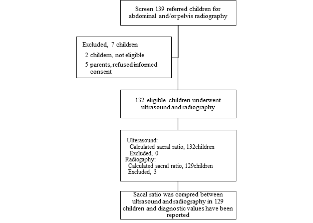
Figure 1: The flowchart of the study
The mean of age in studied children was 52.7±42.5 days. So 82(63.6%) children were male and 47(36.4%) were female. In Table 1, the mean and standard deviation of ultrasound and radiology findings in all infant and separate based on sex are shown. There was a significant difference between male and female in the estimation of Sacrum length(p=0.0002), but no statistically difference between girl and boy in the sacral ratio (p=0.19). Also no statistically difference between male and female in ultrasound finding(P>0.05)
Table 1: Demographics, ultrasound and radiography findings in studied children
|
Variables |
mean |
Male |
Female |
P |
|
Age (day) |
42.37± |
36.09±13.72 |
44.93±10.19 |
0.63 |
|
Sex |
|
82(63.6) |
47(36.4) |
* |
|
Radiography findings: |
||||
|
Sacrum length (mm) |
28.34 ± 4.58 |
27.55±4.4 |
29.22±5.08 |
0.19 |
|
Sacral ratio |
0.70 ± 0.11 |
0.66±0.11 |
0.75±0.1 |
0.002 |
|
Ultrasound findings: |
||||
|
Sacrum length (mm) |
33.19 ± 5.67 |
33.17±4.54 |
34.29±4.38 |
0.35 |
|
Sacral ratio |
0.72 ± 0.05 |
0.72±0.05 |
0.73±0.06 |
0.77 |
According to radiographic finding, the mean of sacrum length was 28.34±4.58 mm and based on ultrasound was 33.19±5.67 mm. The mean of sacral ratio based on radiography was 0.70±0.11 and based on ultrasound was 0.72±0.05 (P-value=0.084).
In Fig. 1, the mean difference size of sacrum was shown in two methods: ultrasound and radiology. Bland Altman chart showed that the mean difference between ultrasonography and radiology was 4.6 mm (confidence intervals of 8.18 ± 5.6), and from 129 infants, only 1 case (0.8%), The size of the sacrum was out of the 95% confidence interval in ultrasound methods.
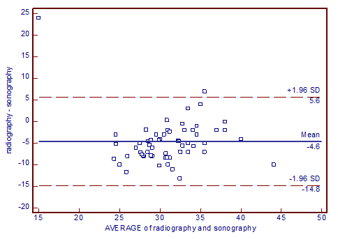
Figure 2: Agreement between radiography and ultrasound for estimation of sacrum length
The mean Sacral ratio in the ultrasound was 0.72 ± 0.086 (range 0,37 - 1.03) and in the radiologic method was 0.70±0.12 (rang: 0.35-1.05). According to Bland Altman's chart (Fig. 2), the mean difference between ultrasound and radiology methods for estimation of sacral ratio was 0.02 (confidence range 0.27 - 0.31). Of the 129 studied patients, in 4 patients (3.1%) sacral ratio were out of 95% confidence interval. According to the above chart, the agreement between ultrasound and radiologic findings was 96.9%.
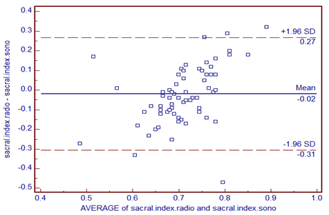
Figure 3: Agreement between radiography and ultrasound for estimation of Sacrum Index
DISCUSSION:
Sacral ratio in newborns that can use for diagnose of sacrum abnormalities is among indications for imaging. Although radiography is considered as the main technique, ultrasonography, that recently developed, with high quality images is considered as an acceptable modality for diagnostic purposes and clinical decision-making, can be considered as an alternative of radiography [14, 15]. In the present study, calculated sacral ratio based on ultrasonography was compared with radiography in infants under six months of age.
The results of our study showed that the use of ultrasonography in determining the sacral index in 97% of the cases was agreed at a 95% confidence level with radiology and the mean difference between the two methods is only 0.02. Therefore, this method can probably be used as a Safe method to use to determine the sacral abnormality. Off course, we were not able to use the ultrasound diagnostic value criteria to determine the abnormalities of sacrum, because all the newborns studied were normal.
This result show that sonographic sacral ratio can be more studied as a good alternative of radiography for sacrum abnormality, however, the small number of samples in our study may be effected our findings and cause to under estimate of ultrasonography method to define sacrum abnormality.
The sacral ratio measurement has been correlated with different diseases and radiography is known as main method of sacral ratio measurement. Both lateral and antero-posterior radiography can used to measurement of sacral ratio but this measurement depends on experience of radiologist and is subjective. Also some other difficulties to measurement of sacral ratio must be considered. First, the radiographs most have high quality, especially in lateral position. Second, the measurement of sacral ratio are based on three single points on the lateral graph and parallel lines on antero-posterior. Third, since the spine may be incompletely ossified in infants younger than 6 months the measured ratio based on graphs may be affected by the infants’ age [9]. The harmful nature of radiation and radiography cost, along with the noted problems, highlighted the importance of the our findings on the use of ultrasound and further research to more clarify the validity of ultrasonography in compare to radiography for the measurement of sacral ratio.
The results of our study showed that ultrasound had a favorable agreement with radiography in the estimation of Sacral index, so that of 129 neonates, in 3.1% of cases, the agreement between the two methods was outside the 95% confidence range.
In one study done by Cyrus et.al. Correlation of Sacral Ratio and Reflux-Related Renal Injury in Children with Vesicoureteral reflux with and without Nephropathy was evaluated. In this study that conducted by Cyrus et al. 200 children under 9 years old with urinary tract infection and according to the VCUG and DMSA results, children with various grades of VUR entered the study. The sacral ratio was measured in the two groups and compared by a radiologist. The case group included 48 males and 52 females with a mean age of 3.7 years old who had reflux nephropathy, and the control group included 47 males and 53 females with a mean age of 4.1 years who did not have reflux nephropathy. Sacral ratio abnormality was detected in 64% and 23.7% [16]. Yadav et al. 140 (83 male and 57 female) adult human sacra were collected from Department of Anatomy, Government Medical College, Aurangabad, Maharashtra. The measurements included ventral straight length and maximum breath of sacrum, sacral index, maximum transverse and antero-posterior diameter and index of body of first sacral vertebra. Demarking points for these parameters were used for identification of sex of sacrum. Results: The average values of sacrum for ventral straight length was found to be 104.7±5.94 mm in male and 92.6±6.1 mm in female, maximum breadth was 102.93±4.83 mm in male and 104.77±6.48 mm in female and sacral index was 98.44±4.69 mm in male and 113.23±5.61 mm in female. The average values of first sacral vertebra for transverse diameter was 48.48±4.21 mm in male and 40.75±3.51 mm in female, antero-posterior diameter was 29.12±2.47 mm in male and 26.93±2 mm in female and index was 60.28±4.96 mm in male and 66.36±5.04 mm in female [17]. In the study of Ahmadi et al. One hundred and twenty children with ARMs were investigated in this study. Sacral ratio (SR) was measured from pelvic X-rays of patients. Among these, 52 patients (43%) had no pelvic X-ray and were excluded from this study. SR was measured by drawing three horizontal lines, through iliac crests (A), tip of coccyx (B) and inferior point of sacroiliac joints (C). The SR was determined by dividing the distance between lines B and C to the distance between lines A and B. In the normal and well developed children, the average ratio is ≥0.74. The SR was higher than 0.70 in 12 (17.7%) children and less than 0.69 in 56 children (82.3%). Among children with ARMs and abnormal SR, 38 cases (68%) had SR of 0.50-0.69; 12 cases (21.5%)had SR of 0.40-0.49 and 6 cases (10.5%) had SR of 0-0.39. In children with ARMs and normal SR, the fecal incontinence was observed in 2 cases (16%). In contrast, 16 cases (29%) with ARMs and abnormal SR had functional disturbance, either fecal incontinence or soiling (P < 0.12). When the patients had an absent sacrum, they had zero possibility for bowel control and frequently had major urinary problems. The sacral feature and SR appear to have a direct influence on the final functional outcome in ARMs. The abnormal SR < 0.7 correlates with poor bowel function. In patients with ARMs, sacral segment and SR are important factors in post operative bowel function disturbance [18]. Lockwood and et al ,To determine the ability of first-year osteopathic medical students to establish sacral base position (SBP) and sacral sulcus depth (SSD), using ultrasonography and to identify the relationship of SBP and SSD to body mass index (BMI) and sex, used ultrasonography to obtain the distance between the skin and the sacral base (the SBP) and the distance between the skin and the tip of the posterior superior iliac spine bilaterally. The SSD (the distance between the tip of the posterior superior iliac spine and the SBP) was calculated. Ultrasound of 211 images were included in the study. The SBP was not significantly different between the left and right sides (36.5 mm vs 36.5 mm; P=.95) but was significantly different between normal and overweight BMI categories (33.0 mm vs 40.0 mm; P94) [19].
To compare the sacral index in a normal pediatric population and in several groups of patients with constipation, anorectal malformations, and vesicoureteral reflux, Caicedo et al, 156 pediatric patients who were classified into four groups (normal, constipation, vesicoureteral reflux and anorectal malformation) were collected . Sacral index values were compared in two projections: frontal and lateral radiographs of the pelvis, obtaining two Key words (MeSH) Vesico-Ureteral Reflux Constipation Sacrum Diagnosis 4303 artículos originales Rev. Colomb. Radiol. 2015; 26(4): 4302-9 measurements. Then, these measurements were compared with international references. The average values of the sacral index for the analysis groups were obtained through quantitative analysis of data for measurements at different projections. In the normal population, the acquired averages for the frontal projections were 0.7022 and 0.7087, and on the lateral view were 0.7257 and 0.7370; for patients with constipation the average value was 0.714 on the frontal projections and 0.7239, and on the lateral views were 0.757 and 0.796. In patients with vesicoureteral reflux, frontal projections showed averages of 0.7117 and 0.7264, and the value on the lateral projection of 0.9708 to 0.9085; for anorectal malformations averages were 0.4618 and 0.4763, being lower when patients had high-type anorectal malformations (0.2893 and 0.3121). Conclusion: The sacral index in patients with vesicoureteral reflux and constipation did not show significant variation compared to the values obtained in the normal pediatric population. In the group with anorectal malformations, these values were below the average when compared with the other groups, with lower measurements in the subgroup of high malformations [20].
In contrast, in a radiographic survey for calculating the Sacral Ratio or estimation of sacral Index, a high quality graph is required to illustrate the sacrum bark landmarks. For this study, two types of anterio posteror and lateral graphs are used. In the lateral alignment aspect, bone components are not clear. In an anterio posterior view, three horizontal lines are used to calculate, while in the lateral view, only three points are used, with three points in comparison to three lines of error. The spine in newborns and children less than 6 months old has not yet been completely bone and may be affected by the X-ray. Also the measures of the Sacral Ratio and Sacral length may not correctly estimated. Therefore, because the aim of radiographic surveys in neonates is screening, in seems that ultrasonography is preferable in determining bone defects. Also Ultrasonography has several potential advantages over radiography. It is portable, less costly and does not expose the infants to ionizing radiation.
CONCLUSION:
Our findings show that sacrum ultrasonography could be safely used in the screening of sacral ratio in infants which lead to reducing the need for radiographs, although, additional assessment are required for accurate assessment of ultrasonography with radiography for sacral ratio assessment to diagnosis of sacrum abnormalities.
Financial support and sponsorship: This study was supported by Isfahan University of Medical Sciences. This design was registered in research faculty of medical school of Isfahan by code 396677
Conflicts of interest: There are no conflicts of interest.
Author's Contribution:
MR: Study idea, study design, interpretation and contribution in writing the paper
MM: Contribution in Data collection and data analysis and preparation of paper
MF: Contribution in interpretation of sonographic and radiologic graphs.
MH: Contribution in Data collection and data analysis and preparation of paper
PCh: Contribution in Data collection.
Author's Contribution:
Maryam Riahinejad: Study idea, study design, interpretation and contribution in writing the paper
Mohsen Masoumi: Contribution in Data collection and data analysis and preparation of paper
Maryam Farghadani: Contribution in interpretation of sonographic and radiologic graphs.
Mehrdadreza hosseinpoor: Contribution in Data collection and data analysis and preparation of paper
Parvaneh Changanian: Contribution in Data collection.
REFERENCES