International Journal of Pharmaceutical and Phytopharmacological Research
ISSN (Print): 2250-1029
ISSN (Online): 2249-6084

Leptadenia pyrotechnica (Forssk.) Decne, a desert plant consumed widely in Saudi Arabia, has many medicinal activities and possible immune effects. Thus, this study determines the immunomodulatory activity of aqueous LP extract on the immune responses and body weight in rats. No published studies on the immune system and body weight effects of aqueous extracts of LP exist. Seventy-two adult Wistar albino rats were equally divided (12 rats per group and equal numbers of females and males) into the innate and adaptive immune responses studies. Each set of groups were orally gavaged with the aqueous extract at a low dose (LD, 20 g/kg body weight) and a high dose (HD, 40 g/kg body weight) while the control was gavaged with 3 ml of water, daily for 21 days. For the innate immune response, the neutrophil adhesion and macrophage phagocytic activity were determined. For the adaptive immune response, the heamagglutinating antibody titer and delayed-type hypersensitivity reaction were measured. Results were compared between the LP groups and the control. The mean daily overall body weight loss, and feed inefficiency ratio for both LP groups were significantly higher, while the mean daily body weight and feed and water consumptions were lower. The LD group had a significantly lower phagocytic index, carbon clearance rate, and neutrophil adhesion percent. The delayed-type hypersensitivity and heamagglutinating antibody titer for the LD group were significantly higher. Thus, consuming the aqueous extract for three weeks led to lower body weight, stimulated adaptive immunity, and suppressed innate immunity.
INTRODUCTION
Overweight and obesity are major problems worldwide with rates increasing steadily [1, 2]. Overweight and obesity lead to a high risk for increased mortality and morbidity. Many diseases and conditions are related to overweight and obesity, such as high inflammation, inflammatory diseases, diabetes, some cancers, cardiovascular diseases, and many others. On the other hand, lower weight and healthy weight lead to lower inflammation and lower rates for inflammatory and other diseases. Therefore, there are many benefits to reducing weight and many people try to reduce weight using natural non-harmful, and non-surgical ways. One common way is by using plants and herbs. The benefits of this approach are the ease of availability and low cost of plants and herbs and, most importantly, the low or no risk of side effects. It is important to use seeds or plants that may lead to a reduction in BW while at the same time they do not lead to any ill effects or toxicity [3]. Some commonly used plants and herbs for reducing BW are ginger, rhubarb, nigella sativa, Cassiae semen, Coptis, and Citrus aurantium [4-6].
Leptadenia pyrotechnica (Forssk.) Decne. (LP) is a desert plant of the Asclepiadaceae family appearing in equatorial areas of Asia and Africa. It is named "markh" in Arabic and "Kip, Khimp and Khip" in other countries, such as Pakistan, Sudan, and India [7]. It is a shrub with many branches that have light green flowers and fruits but rarely any leaves [7]. The plant contains many active compounds, such as cardiac glycosides, alkaloids, flavonoids, tannins, and saponins [8]. Additionally, the stem contains polyphenolic compounds [8, 9], steroids, terpenes, and fatty acids [8]. In Saudi Arabia, LP is added to meats to add flavor and it is eaten as a vegetable. In local folk medicine, LP boiled in water is ingested for the treatment of flu and as a tussive, although there is no scientific evidence for its effects [7, 10]. Other cultures use it to treat tuberculosis [11], rheumatism, and gout, and it is used as an antihistaminic and expectorant [12].
Numerous plants and seeds, such as black seeds, garden cress seeds, soybean, and curcumin, which are considered harmless, have been utilized for thousands of years in traditional and folk medicinal systems across the world to enhance the immune response and for the treatment of many ills [3, 13, 14]. Only one published study [15] investigated the effects of LP on the immune system in rats using the methanolic extract of LP. The immune system is a complex system that is essential for the protection of the body against pathogens. Additionally, the immune system is important for health where many diseases result from its functional failure, such as cancer, arthritis, infectious diseases, inflammation, allergy, and organ failure.
The first line of defense against pathogens is the innate immune response which is mediated through physical and chemical barriers and cells, which include macrophages and neutrophils. Neutrophils play an early role in defending the body against microorganisms during inflammation, where they migrate from blood vessels to the site of infection through intercellular adhesion and with the help of inflammatory signals [16]. ‘Neutrophils’ adhesion to the endothelium occurs through three types of molecules, one of which are the integrins. Integrins are needed for the firm adhesion between the surface of neutrophils and the intercellular adhesion molecule-1 expressed on endothelial cells [17]. The phagocytic monocyte-macrophage system is an important part of the innate immune system involving the blood monocytes that change into the macrophages upon their transfer from the blood circulation into the tissue. In addition, macrophages are characterized by the high production of pro-inflammatory factors, which facilitate the presentation of antigen, the activity of microbicidal, and phagocytic clearance of debris [18].
If the innate immune response fails to destroy pathogens, the adaptive immune response will be activated after approximately four to seven days. Therefore, the adaptive immune response requires a longer time for its activation, but it targets the pathogen with greater precision. Adaptive responses may be mediated by T cells that directly kill pathogens through the cell-mediated immune response, or by B cells that secrete antibodies that enhance phagocytosis through the humoral immune response [19]. Delayed type (or type IV) hypersensitivity is a cell-mediated immune response that results from the direct reaction of antigen-specific memory T cells upon activation by antigen [20]. It develops slowly where the reaction appears after about 18-24 hours, where sensitized T cells release lymphokines, cause macrophage activation and aggregation at the site of antigen invasion, increase vascular permeability, stimulate vasodilation, and initiate inflammation [21].
This study aimed to investigate the effects of the aqueous extracts of LP on the immune system and BW. The aqueous extract was chosen since it is the method of choice in folk medicine and it is the most easily accessible in the home by the general public. This is the first study to examine the effects of the aqueous extract on immunity and BW.
MATERIALS AND METHODS
Preparation of the aqueous LP extract
The young stems of LP were harvested in September 2020, from the Khulais governorate, Makkah, KSA. LP stems were cleaned thoroughly with tap water and a final rinse using distilled water. The aqueous extract was prepared by boiling 500 g of LP stems, in 1 L of water for 5 minutes [11]. The extract was filtered using cotton balls. Subsequently, the extract was allowed to air dry for two days resulting in a semisolid precipitate that was green to brown. The precipitate was stored at 4ºC for about one week.
Experimental animals
Female and male healthy young adult Wistar albino rats, weighing about 170-250 g, were used for this study. The rats were housed in cages with no more than six rats per cage. Cages were maintained at room temperature and exposed to the natural light/dark cycle. All animals were allowed one week to acclimatize to the laboratory conditions before being used for the study. All animals were allowed free access to the standard nutritionally balanced diet and drinking water.
Physiological evaluation for the rats
For physiological evaluation of the rats, mean daily BW, mean overall BW loss, mean percent relative overall BW loss, mean daily feed and water consumptions, and mean daily feed inefficiency ratio (FIR) were determined. For each group, the overall BW loss was determined by subtracting the initial BW from the final BW for each rat. The percent relative overall BW loss was calculated for each rat.
Innate ımmunity assay
For the determination of neutrophils adhesion percent and macrophage phagocytic activity, 36 female and male rats were divided equally into three groups. The ‘two LP’ experimental groups were orally gavaged with the LP extract at a low dose (LD, 20 g LP/kg BW in 3 ml of water) and a high dose (HD, 40 g/kg in 3 ml of water), while the control group was gavaged with 3 ml of water, daily for 21 days. The BW and the feed and water consumptions were measured daily.
Determination of neutrophils adhesion percent
At the end of the experimental period, whole blood was collected in ethylenediamine tetraacetic acid vacutainer test tubes from all rats for the determination of total white blood cells and neutrophils counts (first counts). Subsequently, 1 ml of each blood sample was incubated with 80 mg of nylon fibers (Bon Tool, Gibsonia, USA) at 37°C for 15 min. Then, the total white blood cells and neutrophils were counted a second time. The neutrophils index and percentage of neutrophils adhesion were calculated [22].
Determination of macrophage phagocytic activity
At the end of the experimental period, treatment with LP was stopped for 48 hours. On the 24th day, rats were injected intravenously with 10 µl/g BW of Super Black India ink (Speedball Art Products Company, Statesville, USA) diluted 1:8 times with 0.9% normal saline. Subsequently, 50 µl of blood was withdrawn from each rat at 0 and 15 min after injection and mixed with 4 ml of 0.1% sodium carbonate for red blood cell lyses. The optical density of this solution was measured at 650 nm using a spectrophotometer (Genesys 10SUV-VIS, Thermo Fisher Scientific, Madison, Wisconsin, USA). Finally, rats in each group were sacrificed and the liver and spleen were harvested and weighed. The phagocytic index and carbon clearance rate were calculated [23].
Acquired ımmunity assay
For the determination of haemagglutinating antibody titer and delayed-type hypersensitivity (DTH) reaction, 36 female and male rats were divided equally into three groups as done in the previous section. The rats were administered with LP extract or with plain water daily for 21 days as done above. On the 7th day, rats were injected intraperitoneally with 0.1 ml of goat red blood cells (GRBCs) prepared as described below.
Preparation of goat’s red blood cells
Fresh whole goat blood was drawn from the jugular vein into ethylenediamine tetraacetic acid vacutainer test tubes. Subsequently, the blood sample was mixed with the Alsever’s solution at a ratio of 1:1 and then stored at 4ºC for two weeks.
To prepare the stored blood samples for use in the rats and to isolate the GRBCs, the blood sample was washed three times with 0.9% normal saline and centrifuged for 10 min at 2000 rpm after each wash. Finally, the RBCs were counted by using an Automated Hematology Analyzer (Advia 120, Simens Company, Munich, Germany). The concentration of GRBCs was 2 x 10⁷ cells/ µl.
Determination of haemagglutinating antibody titer
On the 21st ‘day’ of the experimental period, blood samples were collected from all rats into vacutainer test tubes coated with silica and containing polymer gel for serum separation. To determine the haemagglutinating antibody titer [24], 25 μl of each serum sample were serially diluted two-fold in a 96-well V-bottom microtitration plate using 0.9% normal saline. Subsequently, 25 μl of 10% GRBCs in 0.9% normal saline was added to each of the 96-wells and mixed gently. Finally, the microtitration plates were incubated for 2 h at 37oC and visually examined for the phenotype of agglutination.
Determination of delayed-type hypersensitivity reaction
On the 21st day of the experimental period, the rats of all groups were injected with 0.05 ml of GRBCs (2 x 10⁷ cells/ µl) into the right hind footpad. The foot thicknesses were measured before the injection and after 24, 48, and 72 h of the time of injection, using a micrometer screw gauge. The difference between pre-injection and post-injection hind footpad puffiness in mm and footpad puffiness percentage were considered as an indication of the DTH reaction to the particular antigen used (GRBCs) [25].
Statistical analysis
The MegaStat (Version 9.4, Butler University, Indianapolis, Indiana, USA) statistical program was used for the analysis of data. The data were expressed as mean ± standard deviation (SD). The pairwise t-test was used for the significance testing between groups for the normally distributed parameters while the Mann-Whitney test was used for the non-normally distributed parameters. The statistical difference was considered significant for P < 0.05, highly significant for P < 0.01 and non-significant for P ≥ 0.05.
RESULTS AND DISCUSSION
Daily physiological evaluation
The effects of the LP extract on the mean daily BW, overall BW loss, daily feed and water consumptions, and daily FIR compared with the respective control groups for the innate immunity response groups are shown in Table 1. It was found that there were no significant differences in the mean daily BWs between the groups. While, the mean overall BW loss, percent relative overall BW loss and mean daily FIR for the rats in the LD and HD groups were highly significantly higher compared to the respective control. The mean daily feed and water consumptions were highly significantly lower compared with the respective control for the LD and HD groups. For all the above parameters, there were no significant differences between the LD and HD groups.
Results in Table 2 show the mean daily BW, overall BW loss, daily feed and water consumption, and daily FIR for the adaptive immune response study groups as compared with the respective control groups. It was found that the mean daily BW for the rats in the LD and HD groups were highly significantly lower while there was no significant difference between the LD and HD groups. The mean overall BW loss, percent relative overall BW loss and mean daily FIR for the rats in the LD and HD groups were significantly higher and highly significantly higher, respectively, comparing each with its respective control. In addition, compared to the LD group, the HD group was highly significantly higher for the mean overall BW loss and daily FIR while it was significantly higher for the mean percent relative overall BW loss. The mean daily feed consumptions for the LD and HD groups were highly significantly lower compared with the respective control, while the consumption for the HD group was highly significantly lower than for the LD group. The mean daily water consumptions for the LD and HD groups were highly significantly lower compared with the respective control, but there was no significant difference between the LD and HD groups.
Table 1. Daily physiological evaluation for the innate immune response study groups
|
Parameter |
Group |
Mean ± SD |
P-valuea |
P-valueb |
|
Daily body weight * (g) |
Control LD HD |
221.27 ± 17.80 212.35 ± 23.78 205.20 ± 22.94 |
0.320(NS) 0.078(NS) |
0.424(NS) |
|
Overall body weight loss** (g) |
Control LD HD |
0.00 ± 0.00 18.41 ± 16.81 20.50 ± 17.32 |
0.000(HS) 0.000(HS) |
0.682(NS) |
|
Percent relative overall body weight loss** (%) |
Control LD HD |
0.00 ± 0.00 8.33 ± 7.74 9.36 ± 9.36 |
0.000(HS) 0.000(HS) |
0.671(NS) |
|
Daily feed consumption* (g) |
Control LD HD |
118.08 ± 17.22 85.11 ± 9.11 86.91 ± 12.08 |
0.000(HS) 0.000(HS) |
0.566(NS) |
|
Daily water consumption* (ml) |
Control LD HD |
149.40 ± 21.63 122.30 ± 21.46 123.20 ± 25.05 |
0.000(HS) 0.000(HS) |
0.564(NS) |
|
Daily feed inefficiency** ratio |
Control LD HD |
0.00 ± 0.00 1.38 ± 1.25 1.47 ± 0.88 |
0.000(HS) 0.000(HS) |
0.809(NS) |
*Pairwise t-test and** Mann-Whitney test were used for the significance testing
a: Difference between the groups and the control; b: Difference between LD and HD groups
LD: Low dose, HD: High dose, S: Significant, HS: Highly significant, NS: Non-significant
Table 2. Daily physiological evaluation for the adaptive immune response study groups
|
Parameter |
Group |
Mean ± SD |
P-value a |
P-value b |
|
Daily body weight* (g) |
Control LD HD |
246.32 ± 20.48 219.17 ± 16.29 214.73 ± 18.77 |
0.008(HS) 0.002(HS) |
0.637(NS) |
|
Total body weight loss ** (g) |
Control LD HD |
1.25 ± 2.05 10.00 ± 7.86 20.62 ± 9.84 |
0.024(S) 0.000(HS) |
0.008(HS) |
|
Percent relative overall body weight loss** (%) |
Control LD HD |
0.38 ± 0.74 4.13 ± 3.56 8.63 ± 4.24 |
0.023(S) 0.000(HS) |
0.011(S) |
|
Daily feed intake* (g) |
Control LD HD |
196.78 ± 18.73 156.67 ± 34.39 126.65 ± 26.44 |
0.000(HS) 0.000(HS) |
0.001(HS) |
|
Daily water intake* (ml) |
Control LD HD |
243.10 ± 18.52 193.80 ± 48.29 181.30 ± 24.46 |
0.000(HS) 0.000(HS) |
0.290(NS) |
|
Daily feed inefficiency ratio* |
Control LD HD |
0.02 ± 0.02 0.41 ± 0.35 0.98 ± 0.52 |
0.027(S) 0.000(HS) |
0.005(HS) |
*Pairwise t-test and ** Mann-Whitney test were used for the significance testing
a: Difference between the groups and the control; b: Difference between LD and HD groups
LD: Low dose, HD: High dose, S: Significant, HS: Highly significant, NS: Non-significant
Innate ımmune response
Neutrophils adhesion percent
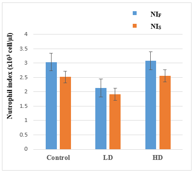
After 21 days of administering the LP aqueous extract, neutrophils adhesion to nylon fibers was measured to assess the functionality of the innate immune response, which is regulated by neutrophils. The mean percent neutrophils adhesion to nylon fibers (Table 3) for the LD group was highly significantly lower while for the HD group it was not different when comparing each with the adhesion for the control group. While the mean percent neutrophil adhesion for the HD group was significantly higher than for the LD group. In addition, the neutrophils index (Figure 1) for the LD group was significantly lower and the HD was not significantly different compared to the index for the control group.
Macrophage phagocytic activity
The effect of the LP aqueous extract doses on the phagocytic activity of macrophages was measured by the carbon removal rate from the blood stream, as shown in Table 3. The LD group showed a significantly lower rate of carbon clearance and phagocytic index when compared with the control group. However, the HD group did not show any significant differences in the rate of carbon clearance and phagocytic index when compared with the control group. Finally, for the comparison between the LD and HD groups for the above parameters, only the rate of carbon clearance showed a significant difference where the HD group was higher.
|
|
|
Figure 1. The effect of the LP aqueous extract on the neutrophils index. Where NIF: Neutrophils index for the first count and NIS: Neutrophils index for the second count |
Table 3. The effect of the LP aqueous extract on the percent neutrophils adhesion, carbon clearance rate, and phagocytic index
|
Parameter |
Group |
Mean ± SD |
P-valuea |
P-valueb |
|
Neutrophils adhesion (%) |
Control LD HD |
19.56 ± 7.37 11.43 ± 5.53 18.11 ± 7.04 |
0.003(HS) 0.600(NS) |
0.033(S) |
|
The carbon clearance rate |
Control LD HD |
0.034 ± 0.02 0.016 ± 0.01 0.034 ± 0.027 |
0.013(S) 0.929(NS) |
0.010(S) |
|
Phagocytic index |
Control LD HD |
8.56 ± 3.44 5.87 ± 1.28 7.02 ± 2.39 |
0.040(S) 0.252(NS) |
0.357(NS) |
Pairwise t-test was used for the significance testing
a Difference between the groups and the control; b Difference between LD and HD
LD: Low dose, HD: High dose, S: Significant, HS: Highly significant, NS: Non-significant
Adaptive ımmune response towards goat red blood cells
Haemagglutinating antibody titer
The haemagglutinating antibody titer test was used to evaluate the effect of the LP aqueous extract doses on the adaptive immune response to the GRBCs, as shown in Table 4. The LD group showed a significantly higher haemagglutinating antibody titer when compared with the titer for the control group. However, the HD group did not show any significant differences in antibody titer when compared with the control and LD groups.
Delayed type hypersensitivity reaction
The DTH reaction induced by the GRBCs was used to evaluate the effect of the LP aqueous doses extract on the adaptive immune response. Compared with the control group, the mean footpad puffiness and percent footpad puffiness were highly significantly higher after 24 h and significantly higher after 27 h and 48 h, respectively, after the injection of the GRBCs into the right footpads for rats in the LD group. However, the rats in the HD group did not show any significant differences in the mean footpad puffiness and percent footpad puffiness after 24, 48, and 72 hours of injecting the GRBCs compared with the control group (Table 4). As for the comparisons between the LD and HD groups, there were no significant differences except for the mean footpad puffiness after 24 h for the HD group that was significantly higher compared to the LD group.
Table 4. Effect of LP extract on haemagglutinating antibody titer and DTH reaction induced by GRBCs
|
Parameter |
Group |
Mean ± SD |
P-valuea |
P-valueb |
|
Haemagglutinating antibody titer |
Control LD HD |
2.6 ± 0.92 3.5 ± 0.76 3.3 ± 0.71 |
0.397(S) 0.132(NS) |
0.537(NS) |
|
Puffiness of footpad (mm) after 24 h |
Control LD HD |
0.79 ± 0.41 1.48 ± 0.51 0.94 ± 0.50 |
0.009(HS) 0.538(NS) |
0.035(S) |
|
Puffiness of footpad (mm) after 48 h |
Control LD HD |
0.38 ± 0.19 0.73 ± 0.36 0.45 ± 0.27 |
0.025(S) 0.610(NS) |
0.072(NS) |
|
Puffiness of footpad (mm) after 72 h |
Control LD HD |
0.30 ± 0.15 0.50 ± 0.25 0.36 ± 0.15 |
0.047(S) 0.517(NS) |
0.162(NS) |
|
Puffiness of footpad (%) after 24 h |
Control LD HD |
32.38 ± 21.86 62.75 ± 27.81 40.44 ± 30.98 |
0.036(S) 0.558(NS) |
0.115(NS) |
|
Puffiness of footpad (%) after 48 h |
Control LD HD |
12.60 ± 10.88 33.30 ± 18.84 19.00 ± 13.17 |
0.010(S) 0.394(NS) |
0.065(NS) |
|
Puffiness of foot pad (%) after 72 h |
Control LD HD |
6.90 ± 3.56 14.00 ± 9.07 11.60 ± 6.37 |
0.0461(S) 0.172(NS) |
0.487(NS) |
Pairwise t-test was used for the significance testing
aDifference between the groups and the control; bDifference between LD and HD
LD: Low dose, HD: High dose, S: Significant, HS: Highly significant, NS: Non-significant
This research study determined the effects of LP extract on innate and adaptive immunity parameters. There is only one published study [15] that determined these effects of LP on rats, although they used the methanolic extract of LP. Therefore, the current findings were compared with the previous study of Rasheed et al. and other studies done using the same methods but with other plants. Compounding the problem of the comparison with previous studies is the fact that most studies on laboratory animals and plants use animals with induced medical conditions or diseases and very few use healthy animals.
The results of the effects of LP on the daily physiological evaluation parameters for the innate and adaptive immune response study groups were compared with the respective control groups. The mean daily BW for the innate immunity rats in both groups were non-significantly (P > 0.05) lower. On the other hand, the mean daily BW for the adaptive immune response LD and HD groups were significantly lower (P=0.008 and P=0.002, respectively). The mean overall BW loss (innate: LD and HD P=0.000; adaptive: LD P=0.024 and HD P=0.000), percent relative overall BW loss (innate: LD and HD P=0.000; adaptive: LD P=0.023 and HD P=0.000), and mean daily FIR (innate: LD and HD P=0.000: adaptive: LD P=0.023 and HD P=0.000), for the groups were significantly higher. The mean daily feed and water consumptions for the LD and HD groups were significantly lower (all P=0.000). Therefore, both doses of the LP extract resulted in higher overall and percent relative BW losses for the study groups, which was linked with lower feed and water consumptions and higher FIR.
There are no previous studies on the effects of LP on BW, thus the current results are compared with those of other medicinal plants and seeds. The current results are in agreement with the previous finding in rats of lower BW upon consumption of an aqueous Lepidium sativum extract [26], Cinnamomum cassia powder [27], and the ethanol and ethyl acetate extracts of Maerua psuedopetalosa [28], compared to controls. Also in agreement are the findings in rats of lower feed and water consumptions using ground Lepidium sativum seeds [14], feed intake using aqueous Lepidium sativum extract [26], and feed intake and food efficiency ratio using Cinnamomum cassia powder [27], compared to the controls.
In contradiction with the current results, previous studies showed an increase in BW of rats using puncturevine plant extract [29], aqueous extract of lepidium sativum seeds in mice [30], and the methanolic extract of ganoderma lucidum in rats [31], comparing each with its control. Also in disagreement with the current findings is the higher percent overall BW gain found in rats consuming ground Lepidium sativum seeds [14].
After 21 days of administering the LP aqueous extract, neutrophils adhesion to nylon fibers was measured to assess the functionality of the innate immune response, which is regulated by neutrophils. The neutrophils adhesion stimulates the process of neutrophils margination in blood vessels and subsequent clearance of the pathogen. The neutrophils index was lower for the LD group than for the control. In addition, the percent of neutrophils adhesion to nylon fibers in the LD group was significantly lower (P=0.003) compared with the same for the control group. This reduction in the adhesion of neutrophils may be due to the downregulation of integrins, which mediate firm adhesion to nylon fibers [17]. However, the HD group did not show any significant differences for the neutrophils index and percent of neutrophils adhesion compared with the respective control. The present findings agree with a study [32] on hydroalcoholic extracts of Cassia occidentalis leaves in healthy rats, which showed a significant reduction in the percentage of neutrophils adhesion. On the other hand, the current results are in disagreement with the previous studies showing a higher percentage of neutrophils adhesion in healthy rats administered with the methanolic leaf extract of Moringa oleifera [33] or the aqueous methanolic LP extract [15] and in mice administered the extract of zapoteca portoricensis [34].
The effect of the aqueous LP extract doses on the phagocytic monocyte-macrophage system was investigated. Macrophages play an important role in the removal of carbon particles from the bloodstream. The LD group showed a significant reduction in the rate of carbon clearance and phagocytic index (P=0.013 and P=0.040, respectively) when compared with the control group. However, the HD group did not show any significant differences in the rate of carbon clearance and phagocytic index when compared with the control group. The lower carbon clearance and phagocytic index imply that this dose of aqueous LP extract has inhibitory properties on the carbon clearance in the blood and the macrophage phagocytic activity. This may be due to our unpublished results of lower mean monocytes count for the LD group, but not the HD group, compared to the control. This lower count of monocytes may lead to lower counts of tissue macrophages since they are interrelated. The current results are in disagreement with previous studies on rats that show significantly higher phagocytic index using the water and petroleum ether extracts of thaumatococcus danielli leaf [35], plectranthus leaves extract [36], and aqueous methanolic LP extract [15]. The present findings agree with a study [37] on the ethanolic extract of Moringa oleifera seeds in healthy mice, which showed a significant reduction of macrophage phagocytic activity.
The haemagglutinating antibody titer test was used to evaluate the effects of the aqueous LP extract doses on the adaptive immune response against GRBCs. The LD group showed a significantly higher (P=0.397) haemagglutinating antibody titer when compared with the control group, implying that the level of circulating antibodies is higher for the LD group in order to bind with the GRBCs to assist in their elimination. However, the HD group did not show any significant differences in antibody titer when compared with the respective titer for the control group and the LD group. This enhancement in the adaptive immune response against GRBCs by the LD may be due to an increase in the functional efficiency and the counts of lymphocytes that are involved in the synthesis of antibodies. To give more proof to this finding is the unpublished finding of higher lymphocytes count for the LD group, but not for the HD group, compared with the count for the control. The present findings agree with the previous studies on healthy rats administered with methanolic leaf extract of Moringa oleifera, [33] and the concoction of Coriandrum sativum and Coscinium fenestratum [38], and on mice using an extract of zapoteca portoricensis [34] which showed a significantly higher mean haemagglutinating antibody titer to sheep red blood cells. The current results are in disagreement with a previous study [39] in healthy mice administered with the aqueous extract of Stachytarpetha jamaicensis, which resulted in significantly higher haemagglutinating antibody titer compared with the control group.
The effects of the aqueous LP extract doses on the DTH reaction induced by the GRBCs were used to evaluate the adaptive immune response. This reaction results from the activation of GRBCs antigen-specific memory T-cells that release lymphokines that lead to the activation of macrophages and their subsequent aggregation at the site of injection. This aggregation results in the increase of vascular permeability, which stimulates vasodilation and the occurrence of inflammation [21]. Compared with the control group, the mean and percent footpad puffiness for the LD group were significantly higher (P=0.009 and P=0.036, respectively) after 24 h, (P=0.025 and P=0.010, respectively) 48 h and (P=0.047 and P=0.0461, respectively) 72 h after the injection. However, the rats in the HD group did not show any significant differences in the mean and percent footpad puffiness after 24, 48, and 72 h of the injection, compared with the control group. As for the comparisons between the LD and HD groups for the mean and percent footpad puffiness, there were no significant differences except for the mean footpad puffiness after 24 h for the HD group that was significantly higher (P=0.035) compared to that of the LD group. Therefore, the LD group rats showed the presence of the DTH reaction in response to GRBCs acting as an antigen, which shows the stimulatory influence of LP on T cells. The present findings agree with the previous studies using the methanolic leaf extract of Moringa oleifera on rats [33], the aqueous methanolic LP extract on rats [15], and the extract of zapoteca portoricensis in mice [34] that showed a higher mean percentage of footpad puffiness compared with the respective controls. On the other hand, the current results are in disagreement with a previous study [39] in healthy mice administered with water extract of Stachytarpetha jamaicensis, which showed a lower mean hind footpad thickness compared to the control group.
Both the LD and HD of the aqueous LP extracts led to lower mean BW and feed and water intakes. The LD of the aqueous LP extract gavaged for 21 days resulted in lower carbon clearance, phagocytic index, and neutrophils adhesion percent, which means that the LD of LP suppressed the innate immune response. In addition, the LD resulted in a higher haemagglutinating antibody titer and DTH reaction (shown by higher footpad thickness), which means that the LD of LP enhanced the adaptive immune response.
CONCLUSION
In conclusion, the LD of the aqueous LP extract affected both the innate and adaptive immune responses, suppressing the innate immune response and enhancing the adaptive immune response. On the other hand, the HD had no such effects. Both doses resulted in lower BW. Thus, it may be advantageous to consume the aqueous LP extract for 21 days for weight loss and its immunostimulant potential for the adaptive immune response and immunosuppress potential for the innate immune response.
Acknowledgments: None
Conflict of interest: None
Financial support: None
Ethics statement: Laboratory animals were treated according to the internationally approved guidelines.