International Journal of Pharmaceutical and Phytopharmacological Research
ISSN (Print): 2250-1029
ISSN (Online): 2249-6084

Nanoparticles have risen rapidly to be one of the superior research scopes. The most suitable in the manufacture of nanoparticles as an alternative to the traditional physical and chemical methods are biological methods, bacteria provide many advantages in this context. The silver nanoparticles show interesting with compared to other salts due to widest applications in medical, therapeutics, and cosmetics. Reports of silver nanoparticle activity as antimicrobials are numerous, but the search on activity against the plant viruses was unpretentious. Diverse leave cultivars of tomato plants (Lycopersicon esculentum L) infected by Cucumber Mosaic Virus (CMV) are showing yellowing leaves, mottling, leaf curl, and leaf shoestring as a result of deformation. To better understand, the antiviral properties of silver nanoparticles are the efficacy of CMV disease control strategies. In this article, we will look at the effective role of silver nanoparticles as an excellent antiviral to CMV isolated from naturally infected tomato plants. Viral identity well confirms by direct ELISA test with antiserum to CMV, TMV, TEV, PVY, TSWV, PMMoV, and ToMV, host range, electron microscopy, and (RT-PCR) studies. The present study will evaluate the effects of exogenously applied silver nanoparticles on the percentage of CMV infection, disease severity, virus concentration, main photosynthetic pigments Phenolic compounds, and total protein and protein profile.
INTRODUCTION
Solanum lycopersicon L. owned by many plants is called the Solanaceae. It is the most popular vegetable throughout the world and the most important commercially grown vegetable in the Mecca regions. In the last years, the tomato plant has been productively used as a model plant to examine the initiation of defense pathways after infected viral bacterial, fungal, and biotic stress [1].
Tomatoes are infected by several viruses, that is, TMV, cucumber CMV [2], PVY [3], TEV [4], TSWV [5], and PMMoV [6]. The CMV virus follows the Bromoviridae family, and it follows the genus Cucumovirus. The virus has a wide range, [7] and can cause many symptoms of infection, for example, yellowing, mosaic, stunting, and deformation, thus causing serious economic losses [8-10].
The attention of scientists in various fields has been attracted about assessing nanoparticles to be one of the most important particles of modern science [11, 12]. There are many ways to produce nanoparticles, such as chemical, physical, and mixed methods, and these methods are quick to produce, but are unsafe and have unwanted residues, and are very expensive, and therefore scientists have tended to create an environmentally friendly and inexpensive way to produce nanoparticles [13-15]. Many scientists in the recent period have made great efforts to take advantage of microorganisms as factories to create environmentally friendly nanoparticles [16]. Scientists have used many eukaryotic microorganisms such as (yeasts, fungi, and plants) and prokaryotes such as (actinomyces and bacteria) in the synthesis of nanoparticles. Silver nanoparticles have been used among many minerals as antiviral, antifungal, antibacterial, and current anticancer agents [17]. In this article, we hypothesize that the discovery of new silver reducing strains, the suppression activity against the plant viruses, and the use of charge-free or charge-credit production route, may lead to the greater possibility for economical, eco-friendly silver nanoparticles production method and inhibit the viral infection caused by CMV infecting tomato plants through virus isolation and identification. In addition, studying the effect of different silver nanoparticles antiviral concentration will collect from different prokaryotic organisms on CMV and evaluate the effects of exogenous applied silver nanoparticles synthesized from different prokaryotic organisms on the infection percentage of CMV, the severity of the disease, concentration virus, the concentrations of the photosynthetic pigments content, Phenolic compounds, and protein components.
We can summarize the roles of silver nanoparticles synthesized from different prokaryotic organisms in the effect on natural and artificial infected tomato plants by CMV or protect healthy profitable tomato failed as listed below:
MATERIALS AND METHODS
Isolation and identification of CMV
Naturally infected tomatoes (Lycopersicon esculentum) with the CMV that develop typical symptoms on upper and lower leaves were collected from Mecca regions.
Virus isolation and propagation
Infected leaves of Lycopersicon esculentum showing a typical symptom suspected to be CMV infections were collected from Mecca regions. The collected leaves samples were serologically checked for CMV infection by DAS-ELISA test as explained by [18] against the following viruses: CMV, ToMV, and TMV. All infected samples were tested in triplicate. Samples with positive reactions were used as a source of CMV.
CMV was isolated and biologically purified from naturally infected tomato plants by on C. amaranticolor Coste and Reyn plants produced necrotic local legions surrounded with little halo edge. It was propagated in healthy Squash Cucurbita pepo seedling, cv. (Vegetable morrow white bush) which reacted as were vein clearing, mosaic, blasters, and leaf narrow [19].
Mechanical transmission of CMV
The virus was transmitted mechanically to healthy Squash Cucurbita pepo seedling, cv. (Vegetable morrow white bush) for virus propagation and preserved under controlled conditions after inoculation for three weeks. The presence of CMV was appropriate with the ELISA test (Enzyme-linked Immunosorbent Assay), and the samples that gave a positive result for the ELISA test were used as a source of the virus in the post. The infested sap was obtained by grinding the leaves of zucchini with a buffered phosphate buffer at a pH7.
Electron microscope
Healthy and infected mesophyll tissue was pieced, fixed, and stained for ultra-thin sectioning according to the standard procedures of [20, 21]. The ultra-thin section was viewed with an electron microscope Unit (JOEL-JEA100 CX).
DAS-ELISA
We examined all samples by using DAS-ELISA with a specific kit of CMV antibodies at 405 nm and was determined by using an ELISA plate reader (Model 550; Bio-Rad, Hercules, CA, USA) [22].
Total RNA extraction and RT-PCR
We extracted total RNA from un-inoculated and inoculated leaves of tomato by EZ-10 Spin Column Total TNA Minipreps Super Kit (BIO BASIC INC) specific CMV1 oligonucleotide (5´GCC/GTAA/GCT/GGA/TGG/AC/AA3´). CMV2 (5`TAT/GAT/AAG/AAG/CTT/GTT/TCG/CG 3`) primers [23], with one-step RT-PCR (Reverse Transcription Polymerase Chain Reaction) amplification of CMV coat protein gene.
Collection of silver nanoparticles synthesized from different prokaryotic organisms
Isolation and characterization of bacteria
The bacterial strains were isolated, cultured and the morphological and physiological characterization of the isolates were performed according to the methods described by [24-26].
Production of biomass
For product biosynthesis of bacterial strains in a nutrient broth medium, they were cultured and incubated on an orbital shaker at 27°C and agitated at 220 rpm. After 24 hours, biomass was harvested and centrifuged for 10 minutes at 5000 rpm. and collected the supernatant material for nanoparticle synthesis.
Silver nanoparticles synthesis
The culture supernatant was added separately to reaction vessels containing silver nitrate at concentrations of 0.5 mM, 1 mM, and 2 mM. The reaction between this supernatant and silver ions was carried out for 24 hours in dark conditions. Same time: The control was also triggered without silver ions along with the experiment flasks [24, 27].
Characterization of silver nanoparticles
Primary detection of silver nanoparticles after 24 h of incubation of the supernatant and silver ions, was done by changing the color of the filtrate. Then, these samples were subjected to optical measurements, which by using a UV-Vis spectrophotometer, were performed. The Fourier Transform Infrared (FTIR) analyzed the interaction between silver nanoparticles and protein.
Using an X-ray diffractometer (Philips PW 1710), the X-ray Diffraction (XRD) checked the silver nanoparticles formation. Under sunlight, the supernatant treated with silver nitrate was evaporated to dryness. Using the Debye-Sherrer formula, the air-dried biomass was analyzed [28].
Inhibitory effects of different silver nanoparticles synthesized by some bacteria on cucumber mosaic virus infecting tomato plants
Material of plants and treatments
After surface sterilization, seeds of tomato susceptible cultivar, cv. Castle rock was germinated at 26 ºC in an incubator on moistened filter paper in sterile Petri dishes for 2–3 days and after one day from germination were transferred to sterilized soil-filled pots kept in a growth chamber. CMV was prepared from fresh severely infected leaves of Tomato Cv. Castle rock and used in these experiments. Plants of similar size were selected and divided into three groups after 21 days of growth. Each group consists of eight treatments from synthesized AgNPs. The names of the treatments were as follows, T1: (Healthy, negative control), T2: (Healthy & NPs1), T3: (Healthy & NPs2), T4: (Healthy & NPs3), T5: (Infected, positive control), T6: (Infected & NPs1), T7: (Infected & NPs2), T8: (Infected & NPs3). Each treatment contains 3 replicates (a replicate is one pot containing three healthy seedlings). The names of the groups were as follows, group 1: Inoculation by the virus with NPs, group 2: spray by NPs before 72 hours from virus inoculation, group 3: spray by NPs after 24 hours from virus infection.
Evaluation of changes in virus infection treated with CMV and NPs treatment
The plant leaves were sprayed with 0.1 μg/μl AgNPs including every part from the leaves. The infection percentage and severity of disease were recorded after three weeks from inoculation, according to the following scale: 0 = no symptoms; 1 = crinkling and light mottling; 2 = crinkling and mild mosaic; 3= crinkling, severe mosaic, and size reduction; and 4 = deformation. Disease Severity (DS) values were calculated using the following formula according to [29].
|
|
(1) |
After three weeks from inoculation, the youngest developed leaves from treated healthy and infected tomato plants were collected for analysis of changes.
Effect of silver nanoparticles on physiological parameter
Determination of photosynthetic pigments
The photosynthetic (chlorophyll a, b and carotenoids) pigments were determined according to [30, 31] in one-half gram of Tomato cv. Castle rock after 21 days from inoculation by (CMV). Leaves were collected and ground in a mortar with 10 ml of 90% acetone, approximately 2-3 gm purified sand, and 2 gm CaCo3 were added to neutralize the acidity of the sap and to prevent the transformation of chlorophyll into pheophytin. The acetone extract was filtered through a center glass funnel of fine porosity (G4) and the remainders were washed with small volumes of acetone 90% until being free from pigments. Each filtrate was made up to 50 ml volume with 85% acetone and Pigment concentrations in [mg g−1 fresh weight (FW)] were calculated using the formula of Lichtenthaler:
|
|
(2) |
|
|
(3) |
|
|
(4) |
|
|
(5) |
Total phenolic content mg/gm (D.Wt.)
A modified Folin-Ciocalteu method [32] was used as shown below. For the determination of total phenols in various leaf samples, 1 ml of ethanolic extracts of each leaf sample was added to 1 ml of 2N Folin-Ciocalteu. Test tubes were heated to about 70 c◦ in a water bath, then left for slow cooling at room temperature. Beak man DK-2 spectrophotometer at a wavelength of 650 nm was used for measured optical densities of these samples.
Protein contents analyses
The tomato cultivar leaves were collected for determining soluble, insoluble and total protein contents of infected and NPs treated leaves compared with control. These methods were according to [33].
Statistical analyses
All data were subjected to analysis of variance (ANOVA) and by two conventional methods of analysis, the means were compared. The SPSS software used in carrying out all statistical tests.
RESULTS AND DISCUSSION
Isolation and identification of CMV
Tomato leaves showing typical symptoms of CMV, i.e., vein clearing, severe mosaic, fern leaf, size reduction, and leaf deformation, were located and collected from the Mecca region.
|
|
|
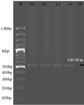
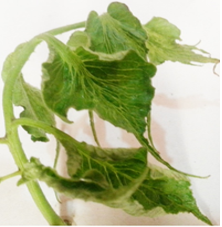
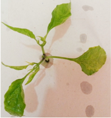
Figure 1. Symptoms caused by natural infection with CMV on tomato leaves; and electropherogram of 1.2% agarose gel from infected tomato with CMV. Electropherogram of 1.2% agarose gel showing PCR amplification from infected tomato with CMV. Lane M, DNA ladder marker, Lanes 1, 2, 3, and 4 represent PCR-positive infected tomato seedling with CMV. Lane 5 represents healthy tomato. |
Samples that yielded a positive reaction in the DAS-ELISA test to CMV were used as a source of virus infection. Thereafter, CMV was isolated and biologically purified from naturally infected tomato plants by a single local lesion on Chenopodium amaranticolor plants that produced necrotic local lesions surrounded with little halo edges. It was propagated in healthy squash seedlings cv. white vegetable marrow bush, which reacted with vein clearing, mosaic, blasters, and leaf narrowing. This result was in agreement with those of [34].
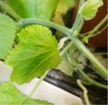
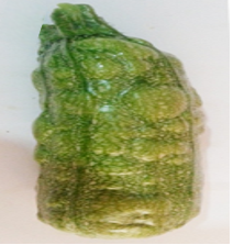
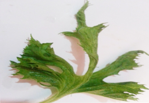
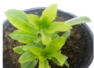
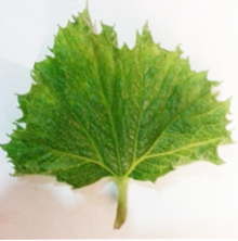
CMV symptoms showing a wide range of discoloration and fruit deformation on the pumpkin (Cucurbita maxima), pepper (Capsicum annuum L.), squash (Cucurbita pepo L.), periwinkle (Catharanthus roseus), and Tomato (Lycopersicon esculentum L.) appeared after 21 days from inoculation with CMV. Symptomatic mosaic appeared on pumpkin; fern leaf, mosaic, and fruit deformation on squash; Yellowing mosaic and leaf deformation on periwinkle; severe mosaic, and leaf deformation on tomato leaf; and mosaic, blasters, leaf deformation, and size reduction on pepper leaves and watermelon leaves Figures (2a, 2b, 2c, 2d, 2e, 2f, and 2g). These results are in agreement with those of [35].
|
|
|
a) |
|
|
|
b) |
|
|
|
c) |
|
|
|
d) |
|
|
|
e) |
|
|
|
f) |
|
|
|
g) |
|
Figure 2. CMV related symptoms show a wide range of fruit malformation and discoloration: (a) Shoestring effect, severe mosaic, leaf deformation on tomato leaf; (b) Symptomatic mosaic on cucumber leaves; (c) Mosaic and fruit malformation on squash; (d) Mosaic, blasters, leaf deformation and size reduction on pepper leaf; (e) Shoestring effect, severe mosaic, leaf deformation on squash leaf; (f) Mosaic, leaf deformation and stunting of plants; (g) Severe mosaic and leaf deformation on pumpkin |
Total RNA extraction and RT-PCR
CMV coat protein gene (CP-gene) was detected in the infected tomatoes using RT-PCR. PCR fragment of the expected size 512 bp was amplified and is shown in Figure 1 [36-38].
Electron microscope
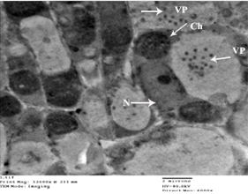
Ultra-structural studies of tomato leaves inoculated with CMV indicated that spherical shaped virus particles associated with cytological changes were found consistently in leaves with symptoms that were absent in symptomless leaves, at the same time, Figure 3 represents the electron micrograph of an ultrathin section of dark green area in infected tomato leaves with CMV, showed the increasing number of chloroplasts with viral particles compared with light green area.
|
|
|
Figure 3. Electron micrograph of an ultra-thin section of light green area in infected tomato leaves with CMV showing few spherical viral particles which were randomly distributed throughout the cytoplasm. Although, the grana and lamellae degeneration and the myelin-like structures’ occurrence was prominent in the chloroplasts; and showing aggregated chloroplasts, swollen chloroplast, amoeboid-shaped chloroplast, chloroplast with stromule, and chloroplast with irregular out-membrane structures such as peripheral vesicle, cytoplasmic invagination, membrane proliferation, and broken envelope. On the other hand, showing deformation in the nucleus shape. Viral particles (VP). Cell organelles (i.e., Ch, chloroplasts; V, vesicles; CW, cell wall; N, nucleus) are also displayed. |
The viral particles occurred mainly in parenchyma cells associated with several vesicles. These vesicles differed in size and were bounded by a single membrane. In addition, the chloroplast from CMV infected tomato was invigilated to include some of the ground cytoplasms of the cell. The nucleus of diseased plants was deformed, as well as viral particles and increasing in the chloroplast in a dark area of leaf tissue. These results are similar to the results obtained by [39].
Bacterial strains identification
The bacterial strains used to stimulate the production of silver nanoparticles with high efficiency by changing the color of the solution from yellow to dark brown and the stability of the brown color of the solution for not less than 48 hours is evidence of the long-term stability of the created nanoparticles. The presence of characteristic Surface Plasmon Resonance (SPR) of silver nanoparticles at 450 nm measured by UV-Visible spectrophotometer was confirmed the AgNPs formation [27].
For identification of B. persicus (KJ743245), and B. licheniformis (KJ743244), BLAST analysis, PCR amplification of the 16S rRNA gene and comparison with known sequences of the GenBank nucleotide database as Bacillus pumilus (KJ743246), were used [27].
The DLS, which was found to be on average 80, 92, and 77 nm, respectively, determines the size distribution of AgNPs synthesized by B. pumilus, B. persicus, and B. licheniformis. Hence, all silver nanoparticles synthesized by the three Bacillus strains were stable at room temperature and they indicated a negative zeta potential [27].
The transmission electron microscope showed that the scale of the nanoparticles is in the range of the nanoparticle scale, most of them being monodispersed with triangular, hexagonal, and spherical shapes [27].
Antiviral activity of silver nanoparticles against CMV
Healthy plants were not affected when exposed to the nanoparticles. Infected plants differ in their reactions when treated with nanoparticles depending on the treatment time (72 hours before infection, with virus infection, and 24 hours after infection). Table 1 show that there is no effect on the concentration of the virus or on the disease severity of tomato plants infected with the virus when treated with nanoparticles 72 hours before viral inoculation.
Table 1. The effect of different silver nanoparticles antiviral on an estimation of the concentration of virus, infection percentage and diseases severity of tomato, cv. Castle rock in the presence of Cucumber mosaic virus under greenhouse condition
|
Percentage of Disease Severity (DS %). |
Percentage of Infection |
* Virus Infectivity |
Virus conc. |
Treatments |
Groups |
||
|
R3 |
R2 |
R1 |
|||||
|
0.00% (0) |
0.00% |
0/3 |
0/3 |
0/3 |
0.034 (-) |
Negative Control |
Group 1 |
|
100% (4) |
100% |
3/3 |
3/3 |
3/3 |
1.153 (+) |
Positive Control |
|
|
8.33% (1) |
33.33% |
1/3 |
1/3 |
1/3 |
0.372 |
Infected & NPs1 |
|
|
44.44% (2) |
88.89% |
3/3 |
3/3 |
2/3 |
0.915 |
Infected & NPs2 |
|
|
5.56% (1) |
22.22% |
1/3 |
0/3 |
1/3 |
0.401 |
Infected & NPs3 |
|
|
0.00% |
0.00% |
0/3 |
0/3 |
0/3 |
0.035 (-) |
Negative Control |
Group 2 |
|
100% (4) |
100% |
3/3 |
3/3 |
3/3 |
1.152 (+) |
Positive Control |
|
|
88.89% (4) |
88.89% |
2/3 |
3/3 |
3/3 |
1.145 |
Infected & NPs1 |
|
|
100% (4) |
100% |
3/3 |
3/3 |
3/3 |
1.163 |
Infected & NPs2 |
|
|
33.33% (2) |
66.67% |
2/3 |
2/3 |
2/3 |
0.994 |
Infected & NPs3 |
|
|
0.00% |
0.00% |
0/9 |
0/9 |
0/9 |
0.035 (-) |
Negative Control |
Group 3 |
|
100% (4) |
100% |
3/3 |
3/3 |
3/3 |
1.153 (+) |
Positive Control |
|
|
8.33% (1) |
33.33% |
1/3 |
1/3 |
1/3 |
0.084 |
Infected & NPs1 |
|
|
22.22% (2) |
44.44% |
1/3 |
2/3 |
1/3 |
0.400 |
Infected & NPs2 |
|
|
5.56% (1) |
22.22% |
0/3 |
1/3 |
1/3 |
0.053 |
Infected & NPs3 |
|
The values are the means (M) of three replicates. The positive and negative control in the Elisa test for virus concentration are 1.492 and 0.113, respectively. Positive control means that infected leaves have typical symptoms and negative control means that infected leaves have no symptoms.
On the other hand, the concentration of virus, the severity of disease, and percentage of infection were remarkably decreased when infected tomato seedling treated with every one of NPs-1, NPs-2, and NPs-3 in the post-infection treatment in which prevented all destructive symptoms caused by the virus.
Systemic severe mosaic, vein clearing, fern leaf, size reduction and leaf deformation yellow mosaic, crinkling, and leave malformation appeared on infected tomato leaves cv. Castle rock by CMV in comparison to healthy leaves. In opposition, silver nanoparticles submission abridged the attendance of destructive symptoms caused by virus progress, particularly when plants spread by silver nanoparticles after 24 hours from inoculation compared with infected and healthy controls, while Mild CMV symptoms were obtained when plants treated by silver nanoparticles with viral inoculums sap.
Silver nanoparticles treatment in Group 1 was recorded a middling velocity or decreased compared with control. Instead, we observed the negative effect of AgNPs treated before 72 hours from inoculation on CMV infection. Ended that silver nanoparticles NPs-3 in Group 3 caused an almost complete reduction of the impact of the virus and viral infection on infected tomato leaves, which led to a reduction in the concentration of the virus and symptoms of infection does not appear on the infected leaves by CMV.
The concentration of CMV, infection percentage, and severity of disease were illustrated in Table 1. Low incidence of symptoms occurred with silver nanoparticles NPs-1, NPs-2 and NPs-3 sprayed after 24 hr of infection and significant decreases in CMV concentration (0.084, 0.400 and 0.053), percentage of infection (33.33%, 44.44% and 22.22%) and disease severity (8.33%, 22.22% and 5.56%) respectively, compared with other treatments. When the silver nanoparticles were sprayed at the same time of infection in Group 1, there was moderate reductions in all symptoms. This suggestion indicates that the silver nanoparticles were affected by the virus during the replication stage inside the cells of infected tomato plants. In contrast, weak to rare reduction occurred in virus concentration (1.145, 1.163 and 0.994), percentage of infection (88.89%,100% and 66.67%) and disease severity (88.89%, 100% and 33.33%), when silver NPs1, NPs2 and NPs3 sprayed pre-viral infection. This may be due to the silver nanoparticles inability to activate the plants’ induced systemic resistance against CMV infection.
Silver nanoparticles may affect the RNA copying during viral multiplication and it is clear that silver nanoparticles significantly influence the inhibition of viral nucleic acid replication when silver nanoparticle particles become less than the size of the particles [17, 40].
Effect of silver nanoparticles on physiological parameter
Determination of photosynthetic pigments
From the data exposed in Table 2, the concentrations of photosynthetic pigments (chlorophyll a, b, total chlorophyll a + b, carotenoids, and Chl a+b / Car) increased in all plants treated with silver nanoparticles, while in infected plants the concentration of the pigment decreased. In addition, these results are in agreement with [27, 41].
Table 2. Changes of photosynthetic pigments in control and cucumber mosaic virus-infected tomato leaves under the effect of different silver nanoparticles antiviral
|
Chl a+b / Car. |
Car. (mg g_1 fresh weight) |
Ch a+b |
Chl b (mg g_1 fresh weight) |
Chl an (mg g_1 fresh weight) |
Treatments |
Groups |
|
3.471± 0.031528 |
0.416± 0.018502 |
1.444± 0.0694718 |
0.398± 0.0346747 |
1.046± 0.020502 |
Negative Control |
Group 1 |
|
2.811± |
0.254± |
0.714± |
0.194± |
0.520± |
Positive Control |
|
|
3.482± 0.016083 |
0.419± 0.017039 |
1.459± 0.03923 |
0.411± 0.033151 |
1.048± 0.033151 |
Infected & NPs1 |
|
|
2.667± 0.056471 |
0.410± 0.016807 |
1.093± 0.037723 |
0.241± 0.015716 |
0.852± 0.016653 |
Infected & NPs2 |
|
|
3.414± 0.027638 |
0.432± 0.043432 |
1.475± 0.024705 |
0.423± 0.032787 |
1.052± 0.011015 |
Infected & NPs3 |
|
|
3.475± 0.015508 |
0.415± 0.063553 |
1.442± 0.014295 |
0.397± 0.037859 |
1.045± 0.020502 |
Negative Control |
Group 2 |
|
2.483± 0.039815 |
0.257± 0.049644 |
0.638± 0.018583 |
0.121± 0.020502 |
0.517± 0.037541 |
Positive Control |
|
|
3.060± 0.026351 |
0.200± 0.01044 |
0.612± 0.015044 |
0.111± 0.0458784 |
0.501± 0.016028 |
Infected & NPs1 |
|
|
3.000± 0.015948 |
0,198± 0.011533 |
0.594± 0.02611 |
0.099± 0.064671 |
0.495± 0.010017 |
Infected & NPs2 |
|
|
2.894± 0.0997807 |
0.348± 0.035774 |
1.007± 0.0351493 |
0.197± 0.048872 |
0.810± 0.0336056 |
Infected & NPs3 |
|
|
3.441± 0.0430039 |
0.420± 0.011547 |
1.445± 0.011547 |
0.399± 0.025774 |
1.046± 0.051316 |
Negative Control |
Group 3 |
|
3.393± 0.0241937 |
0.211± 0.015275 |
0.716± 0.035119 |
0.195± 0.020817 |
0.521± 0.025774 |
Positive Control |
|
|
2.819± 0.0527695 |
0.507± 0.015275 |
1.429± 0.060828 |
0.378± 0.011547 |
1.051± 0.021213 |
Infected & NPs1 |
|
|
3.463± 0.0967720 |
0.413± 0.01 |
1.430± 0.025865 |
0.387± 0.036056 |
1.043± 0.047258 |
Infected & NPs2 |
|
|
2.972± 0.0876603 |
0.508± 0.015678 |
1.510± 0.0623244 |
0.410± 0.032146 |
1.098± 0.049329 |
Infected & NPs3 |
Mean of three replications and ± is the standard deviation
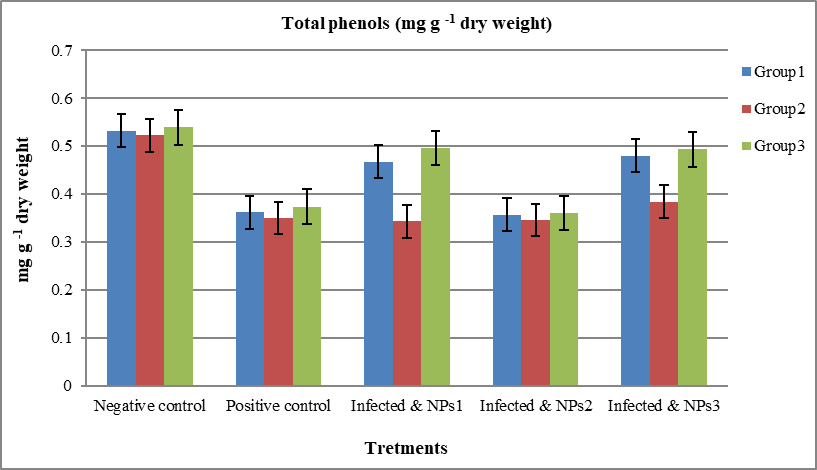
The total phenolics content (mg /g-1 dry weight) reached (0.362, 0.350 and 0.374) in infected tomato leaves cv. Castle rock when inoculated by CMV compared with healthy plants (0.532, 0.523, and 0.539). There was a significant increase in phenolic content at all levels of treatment in tomato leave that were treated with silver nanoparticles (Figure 4a). These results agree with [27, 42]. On the other hand, [43] mentioned that the under-stress condition, including viral infection, stimulation, and increased activity of phenolic play role in defense mechanism.
|
|
|
a) |
|
|
|
b) |
|
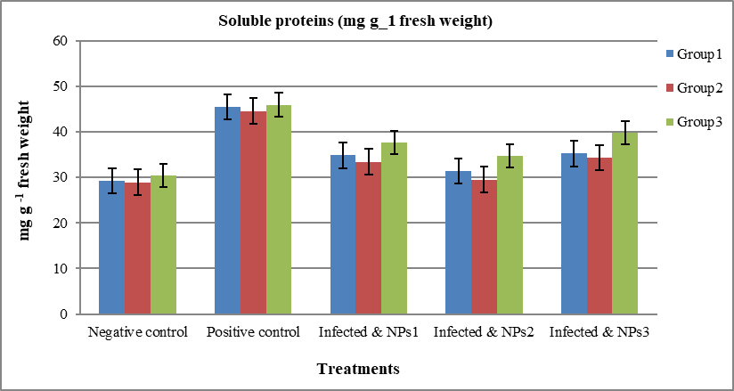
Figure 4. a) Changes of Total Phenols (mg g -1 dry Weight) in Control and Cucumber Mosaic Virus-infected Tomato Leaves under Effect of Different Silver Nanoparticles Antiviral. b) Changes of Soluble Proteins (mg g_1 Fresh Weight) in Control and Cucumber Mosaic Virus-Infected Tomato Leaves under Effect of Different Silver Nanoparticles Antiviral |
All plants treated by silver nanoparticles showed a higher accumulation of phenolic contents compared with infected leaves. [44-47] mentioned that a significant decrease in the alkaloids and phenols contents was observed in the leaves of Datura stramonium inoculated with Potato virus x.
Protein composition and protein patterns analyses
High values from contents of total soluble protein (mg g_1 fresh weight) were recorded in infected tomato plants treated with NPs-1, NPs-2, and NPs-3 after 24 hours from inoculation with CMV (37.64, 34.71, and 39.83), respectively. In comparison with healthy plants (30.41) and other treatment groups shown in Figure 4b. Therefore, all treatments of silver nanoparticles showed lower accumulated total soluble protein content compared with infected leaves, [27, 41, 48, 49].
CONCLUSION
In this study, tomato leaves showing typical symptoms of CMV were located and collected from the Mecca region. CMV was isolated and biologically purified by a single local lesion. CMV symptoms showed a wide range of discoloration and fruit deformation on many host plants and detected the CP-gene of CMV in infected tomatoes by RT-PCR. Ultra-structural studies of tomato leaves inoculated with CMV indicated that spherical-shaped virus particles associated with cytological changes were found consistently in leaves with symptoms. The bacterial strains used to stimulate the production of silver nanoparticles with high efficiency by changing the color of the solution from yellow to dark brown. The presence of characteristic Surface Plasmon Resonance (SPR) of silver nanoparticles at 450 nm measured by UV-Visible spectrophotometer was confirmed the AgNPs formation. The spherical shapes from AgNPs at the nanoscale were confirmed by using TEM (Transmission Electron Microscopy) images. The disease severity of tomato plants infected with the virus was not affected or virus concentration when treated with nanoparticles 72 hours before viral inoculation. Instead, viral concentration, disease severity, and percentage of infection were remarkably decreased when infected tomato seedling treated with every one of NPs-1, NPs-2, and NPs-3 in the post-infection treatment in which prevented all destructive symptoms caused by the virus.
The concentrations of photosynthetic pigments increased in all plants treated with silver nanoparticles, while in infected plants the concentration of the pigment decreased. There was a significant increase in phenolic content at all levels of treatment in tomato leave that were treated with silver nanoparticles. High values of total soluble protein contents were recorded in infected tomato plants treated with AgNPs after 24 hours from inoculation with CMV.
Acknowledgments: We would like to thank the Deanship of Scientific Research (DSR) at the University of Jeddah, Jeddah, Saudi Arabia.
Conflict of interest: None
Financial support: This project was funded by the Deanship of Scientific Research (DSR) at the University of Jeddah, Jeddah, Saudi Arabia., under grant number No. (UJ-02-101-DR). The authors, therefore, acknowledge and appreciate DSR for technical and financial support.
Ethics statement: None