International Journal of Pharmaceutical and Phytopharmacological Research
ISSN (Print): 2250-1029
ISSN (Online): 2249-6084

Green synthesis of silver nanoparticles (AgNPs) was achieved by the bio-reduction of silver nitrate using Walsura trifoliata plant aqueous leaf extract. The AgNPs are characterized by UV-Vis, FT-IR, and TEM. The AgNPs formation was preliminarily confirmed by Ultraviolet-Visible (UV-Vis) spectrophotometer. The synthesized AgNPs in the solution has shown a lesser absorption at 434nm. The Fourier-Transform Infrared (FTIR) Spectroscopy was used to validate the presence of several functional groups responsible for decreasing and stabilizing during the biosynthesis process. The TEM is used to know the morphology, size, and agglomeration pattern of the nanoparticles. The characterization of silver nanoparticles revealed a spherical crystallite nature with particle sizes ranging between 21.08-24.91nm, the average size of the nanoparticles 22.48nm. The synthesized AgNPs showed remarkable antimicrobial activity against clinically isolated two Gram-negative bacteria (K. pneumoniae and E.coli) and two Gram-positive bacteria (B. subtilis and S. aureus); and antioxidant (2, 2-diphenyl-1-picryl-hydrazyl (DPPH), H2O2 and NO radical scavenging activities.
INTRODUCTION
Utilized by human beings for over seven thousand years, silver metal is known as a potent antibacterial agent that has the ability to eliminate several types of microorganisms causing diverse infectious diseases [1]. Nanotechnology is an essential area in the modern sciences, which deals with the synthesis of particles with dimensions that ranges from 1 to 100 nm [2]. This exclusive feature of the nanoparticles provides them with a crucial role in biomedicine and energy field, optic, and other related healthcare applications [3]. Metal nanoparticles have definite characteristics due to their great physicochemical properties, like strong optoelectronic, thermal, and catalytic properties, high surface volume ratio, and ease to synthesise in controllable morphology and distinct crystallinity. Among the nanoparticles, noble metal nanoparticles are known such as gold, silver, and platinum [4]. However, nanoparticles of metal, metal oxides, and metal nitride have the ability to be synthesized by the use of chemicals (viz. precipitation and reduction) synthesis nucleation growth method, physical (sputtering and physical vapour deposition) preparation method, and biologic methods [5, 6]. In the last couple of years, nanoparticles are greatly employed in cosmetics, medicines, and food preservatives that come into direct contact with a human. Especially, metal nanoparticles and metal oxides such as silver, gold, platinum, ruthenium, palladium, iron oxide, rare-earth oxides, and silver nanoparticles are used for antimicrobial activities, drug delivery, and cancer therapy [7-11]. Now these metal nanoparticles utilized in shampoo, soap, cosmetics, and toothpaste as well [12-14]. In the interest of many researchers, numerous nanoparticle applications grabbed across the globe. Silver is one of the metal nanoparticles focused much interest because of its enormous applications [15]. Silver has diverse biological activities such as antimicrobial [16] antihelmintic and wound healing activity [17] antilarvicidic [18] antioxidant [19] anticancer [20] anti-inflammatory [21] hepatoprotective [22]. Previously the nanoparticles can be synthesized by using physical, and chemical approaches. However, the physical and chemical methods are the most expensive, time-consuming, energy consumption, and much more chemicals are required for the production of nanoparticles and they cause hazards to the environment and also unsuitability for biological applications. Instead of the above two methods, researchers need to develop an alternative method. The researchers developed the alternative method biological method to reduce the above-mentioned disadvantages and environmentally safer than the physical and biological methods. This method overcame most of the setbacks by the synthesis of silver nanoparticles through employing several biological agents like algae, bacteria, enzymes, fungi, oligosaccharides, polysaccharides, DNA, and human cell lines [23]. The biological agent's synthesis also has some disadvantages such as bacteria requiring high sterile environments and preservation [24] Moreover, the rate of synthesis is slow and a restricted amount of sizes and shapes are malleable in comparison to the mechanism involved in plant materials [25]. While synthesis of nanoparticles by fungi as a result of feasible handling biomass and economic capability can lead to contamination considering it can cause genetic manipulation of the organism, and also synthesis rate is slow. Accordingly, the utilization of plant extracts is much more useful than microorganisms due to the low cost as we do not need any particular preparation for the culture and isolation techniques and it is easily scaled up for huge-scale synthesis of nanoparticles [3]. Green synthesis has an edge that is environmentally acceptable, as well as economical because it utilizes biocompatible materials for synthesizing silver nanoparticles. Even though the actual procedure of nanoparticles biosynthesis through diverse plant extracts is unclear, it is recommended that the bio-molecules in the plant extracts like protein, phenol, and flavonoids have a magnificent position in reducing the of metal ions and capping the bio-synthesized nanoparticles [26]. The vital role is usually played by the phyto-constituents of the plant, namely, Primary and secondary metabolites, such as sugars, proteins polyphenols, phenolic acids, ketones, terpenoids, amides, etc. Moreover, in the majority of cases, a reducing agent from the plant extract plays a role as both capping and stabilization agents [27]. Currently, synthesis of NPS has been synthesized by the greener method using plant extracts from various plant species victoriously and plant materials act as reducing agents like Copper NPs from Magnolia Kobus [28] Cadmium oxide nanoparticles through Achillea wilhemsii [29] calcium nanoparticles from Boswellia ovalifoliata [30] Gold nanoparticles from Avena sativa [31] Palladium nanoparticles from Cinnamomum camphora [32] Zinc oxide nanoparticles from Catharanthus roseus [33] Silver nanoparticles from Adansonia digitata [34] Iron oxide nanoparticles from Medicago sativa [35].
Walsura trifoliata (A.Juss.) Harms. Belongs to Meliaceae (Syn: Walsura piscidia Roxb., Heynea trifoliata A. Juss). The plant is well-reputed in the traditional system of medicine and is used by tribal peoples to treat diverse diseases like skin allergies, astringents, and diarrhoea [36]. The plant is an ever-green tree distributed widely in the tropical areas of Asia, Southern China, India, Malaysia, and Indonesia [37]. It grows on dry deciduous forests of 200 to 300 m in height. The earlier reports described about the bark of the plant possess stimulant, expectorant, emmenagogue, and emetic properties. The fruit pulp is used as fish poison [38]. The bark extract of the plant exhibited activity against pathogenic microorganisms [36]. But not many laboratory studies made confirming the traditional usage [39]. The present study is focused on characterization and evaluating the antibacterial included antioxidant activity of AgNPs using Walsura trifoliata aqueous leaf extract.
MATERIALS AND METHODS
The collected plant material (leaves) was cleaned by running tap water thrice and followed by distilled water twice. Later all leaves were wiped through tissue paper, then cut into small pieces and dried for up to three weeks under shade dried to evaporate moisture content and made ground fine powder using an electric blender and it was stored in the amber color bottle until further work.
Preparations of bark extract
The leaf powder was sterilized at 121oC for 5 min. 20 g of powder was taken into a 500 ml sterile Erlenmeyer conical flask and 200 ml of sterile Milli-Q water was added and boiled for 30 min at 100oC. Then the leaf extract was collected in a separate conical flask by a standard filtration method. The filtrate was used for characterization, antibacterial and antioxidant activities.
Chemicals and preparation of 1mM Ag (No3)2
10 grams quantity of Silver nitrate was purchased by Hi-media Company and the 1 mM silver nitrate solution was prepared using the sterile Milli-Q water, then this was taken into the amber-colored bottle until the synthesis.
Green synthesis of silver nanoparticles
Synthesis of AgNPs was carried out by the leaf aqueous extract of Walsura trifoliata was added to 1 mM of Ag (No3)2. 10 ml of leaf aqueous extract were taken into a 250 ml sterile conical flask and it was titrated by 100 ml of 1 mM of Ag (No3)2 solution with heating at 800C for 30 min. Later it was centrifuged at 15000 rpm for 15 min to remove the presence of biological admixture, and this was used for characterization and evaluation of its antimicrobial and antioxidant activities.
Characterization of the nanoparticles
Biologically synthesized Silver nanoparticles (Ag NPs) were analyzed using the recent equipment. Ag NPs of Walsura trifoliata were performed by UV-Vis spectrophotometer (Nanodrop contain scan range 190 to 750 nm) to find out which of the nanoparticles interacted in decreasing the nanoparticles by Surface Plasmon Resonance (SPR) mechanism. To understand which phytochemicals were certainly involved in the capping and stabilization of the nanoparticles was performed with the Fourier Transform Infra-Red (FT-IR, ECO ART), Bruker, Ettlingen, Karlsruhe, Germany by KBr pellet method. The Transmission Electron Microscopy (TEM) analysis was done by the HF-3300 advanced 300 kV TEM from Hitachi.
Antibacterial studies
The antibacterial activity was performed by using standard protocol followed by the disc diffusion method [40]. The bacterial strains were procured from the Dept. of Microbiology, Sri Venkateswara University, Tirupati. The aqueous leaf extract of synthesized AgNPs was tested for antibacterial activity against selected two-gram negative like Escherichia coli, Klebsiella pneumoniae, and two-gram positive bacteria such as Bacillus subtilis and Staphylococcus aureus. For this 20µl of plant extract, Ag (NO3)2, Ag NPs, and streptomycin were applied on separate sterile filter paper discs (What man No.1 filter paper disc with 7 mm diameter) and allowed to dry before being placed on nutrient agar medium. The entire assay was done in triplicates and incubated at 370C for 24 hours. The zone diameter was measured in centimeters using the scale and the results were tabulated.
Anti-oxidant activity of WT-agnps
The antioxidant assay of leaf extract silver nanoparticles (AgNPs) revealed significant antioxidant activity.
DPPH radical activity by Bio-synthesized WT-AgNps
The in-vitro anti-oxidant activity of the WT-AgNPs was calculated through the 2, 2’- diphenyl-1- picrylhydrazyl (DPPH) radical scavenging method as described earlier [41]. The spectrophotometric test employed the stable radical DPPH as a reagent. To do this, 1 ml of various concentrations of test samples such as plant extracts and biosynthesized WT AgNPs (25, 50, 75, and 100 µg/ml) in methanol were added to 0.004% (w/v) added to 4 ml of methanol solution of DPPH. Following 30 minutes incubation time at room temperature, we recognize the absorbance (RSA) against blank at 517 nm. The inhibition rate (%) of free radical generation by DPPH was calculated by the following assimilation. The DPPH activity expressed IC50 values. The assay was carried out in triplicates.
|
RSA(%) = [(′ AC′ −′ AS′ )/( ′ AS′ )] × 100 |
(1) |
where ‘AC’ stands for the absorbance of the control reaction (containing all reagents except the test comi.e. the plant extracts and the Dc-AgNPs) and ‘AS’ stands for the absorbance of the test compound, the tests were carried in triplicate.
Nitric oxide scavenging activity
Nitric oxide scavenging activity was measured by slightly amended methods [42]. Nitric oxide radicals (NO) were generated from sodium nitroprusside. Test solution (plant extract and WT AgNPs), incubated at 250° C. for 150 min, and 1 ml of the reaction mixture was treated with 1 ml of Griess reagent (1% sulfanilamide, 2% H3PO4 and 0.1% naphthylethylenediamine dihydrochloride). Absorbance of compounds was measured at 546 nm.
H2O2 scavenging activity
Hydrogen peroxide scavenging (H2O2) efficacy of the test reaction mixtures was ascertained from the literature procedure [43]. The solution of H2O2 (40mM) prepared in phosphate buffer (pH7.4) was added to various concentrations of (25, 50, 75, and 100 µg/ml) the test compounds (like plant extract and WT-AgNPs). 3.4 ml phosphate buffer was added to the H2O2 solution (0.6 ml, 40 mM). The absorbance values of the reaction mixture were written down at 2 30 nm.
RESULTS AND DISCUSSION
UV-Visible spectroscopy analysis of WT-AgNPs
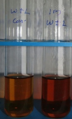
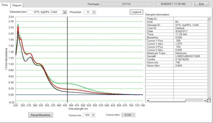
When the aqueous leaf extract of Walsura trifoliata was mixed with the 1mM of Ag(NO3)2 solution, the color visible turned into deep brown from light brown which is the initial technique to corroborate that the synthesized nanoparticles are silver. UV-Vis is the basic parameter; it provides information through exhibited peaks accurately related to specific nanoparticles. The broad peak was acquired at 434 nm (Figure 1a), because of SPR (Surface Plasmon Resonance) of silver nanoparticles in the reaction mixture. The SPR peak divulged numerous morphological features of nanoparticles (NPs), i.e. dispersion, shape, size, and stability of the nanoparticles. The same kind of outcomes was seen in Moringa oleifera leaf mediated synthesis of silver nanoparticles [44].
FT-IR Spectral analysis of WT-AgNPs
Fourier Transform Infrared (FT-IR) of AgNPs was conducted to know the possible biomolecules responsible for capping and stabilization of the nanoparticles and with the help of broad peaks, categorized the biomolecules. For this Fourier Transform Infrared spectrum was analyzed between scan ranges from 4000- 500 cm-1. Here the intensive peaks exhibited at 3365, 2931, 1613, 1409, 1072, 896, 805, 779, 632 and 481 cm-1. The sharp peak 3365 assigned N-H medium stretching primary amine, 2932 assigned to C-H medium stretching Aliphatic group, 1913 belongs to the α,β unsaturated ketone comprising the C=C bond stretching. The broad peak was acquired at 1409 cm-1, it corresponds to =C-H medium bending alkane. The acquired intensive peak at 1072 cm-1 belongs to the primary alcohol, its bond –C-O strong. The assigned peak at 805 cm-1 belongs to the weak Al-Mg-OH deformation. The intensive peak procured at 779 cm-1 corresponds to 123 trisubstituted with containing C-H bending. The peak at 632 cm-1 corresponds to C-S stretching thioethers and 481 cm-1signed –S-S stretching polysulphides (Figure 1b). These all functional groups participated to form nanoparticles and the compounds act as capping and stabilization agents to prevent agglomeration in the reaction mixture. Similar types of results were reported previously by other researchers [45].
|
|
|
|
a) |
b) |
|
Figure 1. UV- Vis and FTIR studies. a) UV- visible absorption at 434 nm of WT-AgNPs, b) Fourier- Transform Infra-Red (FT-IR) spectra of bio-synthesized AgNPs of WT-AgNPs |
|
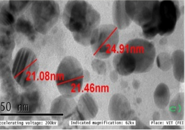
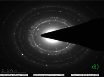
TEM analysis
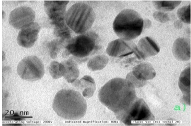
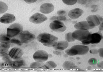
Biosynthesized AgNPs of Walsura trifoliata were characterized by HR-TEM (Transmission Electron Microscopy), which provides micrographs and images at high magnifications (20nm). These higher-resolution micrographs help to find the shape, size, distribution, agglomeration pattern, and surface morphology of the nanoparticles. At 20 nm, higher magnification studies of the biosynthesized Ag NPs revealed very small-sized NPs were seen, size was between 21.08 to 24.91nm owing to spherical, and agglomeration was not observed between the particles. The average size of the nanoparticles was 22.48nm (Figure 2). These types of results were observed in leaves extract of Andrographis serpyllifolia [46].
|
|
|
a) |
|
|
|
b) |
|
|
|
c) |
|
|
|
d) |
|
Figure 2. TEM micrographs of AgNPs of Walsura trifolita. a) At 20 nm no agglomeration found, b) At 50nm AgNPs are spherical in shape, c) At 50 nm average size of the AgNPs is 22.48nm, d) At 51 nm beam of TEM |
Antibacterial studies of AgNPs
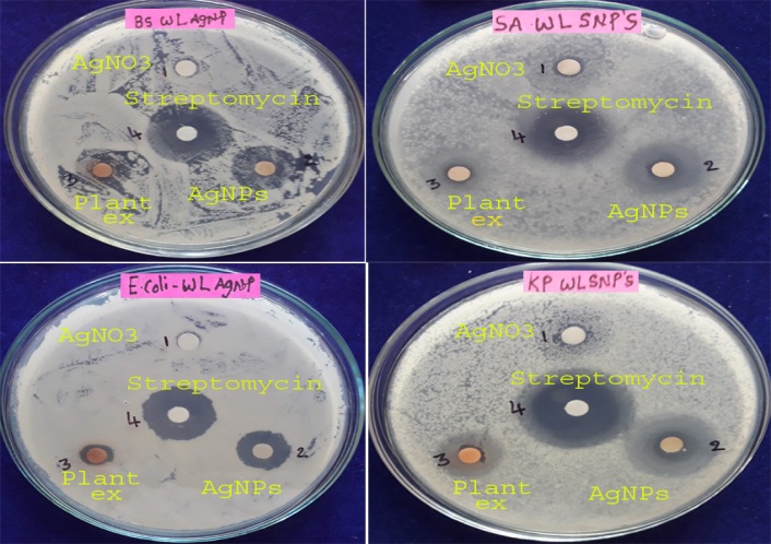
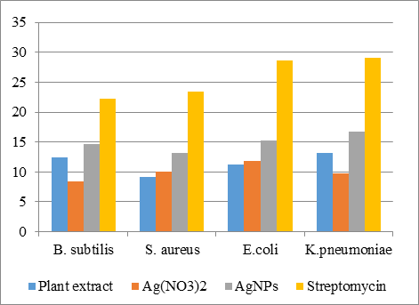
To perceive antimicrobial efficacy of biologically synthesized AgNPs from leaf aqueous source of Walsura trifoliata was analyzed against two-gram positive (B. subtilis- MTTC-441 and S. aureus- MTTC -731) and two-gram negative (E.coli- MTTC-443 and K.pneumoniae- MTTC-741) by disc diffusion assay. The selected plant leaf source AgNPs exhibited excellent antimicrobial activity against Gram-positive (+ve) and Gram-negative (–ve) bacteria. Among the bacterial pathogens, the highest zone of inhibition was seen in K.pneumoniae and E. coli (Figures 3 and 4, Table 1). Antimicrobial activities of AgNPs are reliant on the size and shape of the nanoparticles. The size of the nanoparticles also gives more space to interact with microorganisms. According to the previous reports smaller-sized nanoparticles have maximum antimicrobial activity than larger-sized particles due to they have a large surface to interact with bacteria efficacy which leads to cell death. The present study revealed Gram-negative bacteria showed more susceptibility than Gram-positive bacteria. The Gram-positive bacteria comprise a dense layer made up of peptidoglycans (together with a polypeptide containing proteins) when compared to the Gram-negative bacteria and AgNPs penetration happens easily by the membrane of Gram-negative bacteria and resulting in cell death occurred due to the process involving electrostatic force and endocytosis. Similar kinds of results were observed in leaf-assisted green synthesis of silver nanoparticles from Vitex negundo (Karu Nochchi) Characterization and antimicrobial studies [47].
|
|
|
Figure 3. Antimicrobial activity of biologically synthesized WT-AgNPs against Two gram-positive bacteria and two gram-negative bacteria. 1. Ag solution 2. WT-AgNPs 3.Plant extract 4.Streptomycin |
Table 1. Zone of inhibition (mm) of WT-AgNPs on selected Gram-positive and Gram-negative bacteria with plant extract, Ag(NO3)2, and Streptomycin
|
S.no |
Name of the organism |
Zone of inhibition (mm) |
|||
|
Plant extract |
Ag(NO3)2 |
AgNPs |
Streptomycin |
||
|
1 |
B. subtilis |
12.5±0.088 |
8.4±0.057 |
14.7±0.033 |
22.3±0.066 |
|
2 |
S. aureus |
9.2±0.066 |
10.1±0.11 |
13.2±0.088 |
23.4±0.057 |
|
3 |
E.coli |
11.3±0.033 |
11.9±0.088 |
15.2±1.25 |
28.7±0.066 |
|
4 |
K.pneumoniae |
13.2±0.057 |
9.8±0.066 |
16.8±0.033 |
29.1±0.115 |
|
|
|
Figure 4. Graphical representation of antimicrobial studies using with WT-AgNPs |
Anti-oxidant activity
In this research, we ascertained the free radical scavenging activity, which is measuring the antioxidant rates in aqueous leaf extract and bio-synthesized AgNPs of Walsura trifoliata by diverse methods viz. DPPH, H2O2, and NO antioxidant activity. Plants are usually wealthy in polyphenols.
2, 2- diphenyl-1- picrylhydrazyl anti-oxidant (DPPH) activity
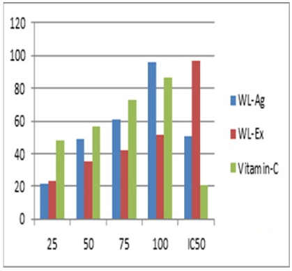
Bio-synthesized AgNPs were determined through the DPPH method. The activity depends on the reduction of DPPH radical from DPPH to DPPH-H, hydrogen donating anti-oxidant. The in-vitro anti-oxidants activity and half maximal inhibitory concentrations (IC50) values of the Walsura trifoliata leaf extract biologically synthesized WT-AgNPs were described in the table (Table 2a and Figure 5a). The revealed results that the DPPH ant-oxidants activity was increased by the concentration of the test samples. Plants possess a wealth of bioactive compounds like flavonoids and tannins relates to the phenolic compounds included different polyphenols, that are a pivotal group of phytochemicals that perform as primary anti-oxidants of free radical scavengers [48]. The highest free radical activity was seen in the WT-AgNPs with 96.2% at 100 µg/mL concentrations while the lowest activity was observed in 21.25% at 25 µg/mL respectively. The DPPH activity showed excellent activity with WT-Ag NPs than a plant extract.
H2O2 scavenging activity
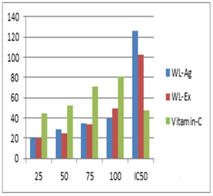
The hydrogen peroxide scavenging activity results exhibited that significant radical scavenging activity was observed by the plant extract and synthesized WT-AgNPs. The highest radical scavenging activity was shown at 39.5% at 100 µg/mL concentrations with the bio-synthesized WT-AgNPs and aqueous leaf extract exhibiting 49% at 100 µg/mL. The lowest activity was observed with WT-AgNPs 20.3 at 25 µg/mL and 19.8 at 25 µg/mL in aqueous leaf extract of the selected plant. In this case, the lower concentration of AgNPs exhibited susceptible H2O2 radical scavenging activity (Table 2b and Figure 5b).
NO antioxidant activity
The nitric oxide scavenging activity of aqueous leaf extract and bio-synthesized AgNPs of Walsura trifoliata detected the highest radical scavenging activity of 45.5% at 100 µg/mL concentrations (Table 2c and Figure 5c). The activity revealed that the lower concentration of AgNPs showed more significance than plant extract of the multipurpose medicinal plant tree taxon.
Table 2. DPPH Anti-oxidant activity of plant extract and WT-AgNPs
|
a). DPPH activity |
|||||
|
Compounds |
25 |
50 |
75 |
100 |
IC50 |
|
WL-Ag |
21.25 |
49 |
60.8 |
96.2 |
51 |
|
WL-Ex |
22.9 |
35.2 |
42.3 |
51.6 |
96.8 |
|
Vitamin-C |
48.4 |
56.5 |
72.8 |
86.6 |
20.6 |
|
b). H2O2 activity |
|||||
|
WL-Ag |
20.3 |
28.2 |
34.2 |
39.5 |
126.5 |
|
WL-Ex |
19.8 |
25.2 |
33.7 |
49 |
102 |
|
Vitamin-C |
44.7 |
52.2 |
70.5 |
80.4 |
47.8 |
|
c). NO activity |
|||||
|
WL-Ag |
19.6 |
23.8 |
37.6 |
45.5 |
109.8 |
|
WL-Ex |
19.4 |
23.6 |
47.3 |
56.7 |
79.2 |
|
Vitamin-C |
46.8 |
53.4 |
74.2 |
83.5 |
46.8 |
|
|
|
a) |
|
|
|
b) |
|
|
|
c) |
|
Figure 5. Graphical representation of antioxidant activity of WT-AgNPs. a) DPPH activity, b) H2O2 activity, c) NO activity |
In conclusion, the results divulged that both plants leaves sourced aqueous extract and biologically synthesized WT-AgNPs possess magnificent anti-oxidant activity. The DPPH activity as a more significant method in comparison with H2O2 and nitric oxide ant-oxidant radical scavenging activity.
CONCLUSION
The Silver Nanoparticles (AgNPs) were synthesized successfully using the aqueous leaf source of Walsura trifoliata. In which reaction leaves aqueous extract was used as a capping agent for the stabilization of the biologically synthesized nanoparticles. For the preparation WT-AgNPs, we followed the simple, cost-effective, eco-friendly approach, a small number of chemicals, and utilized a biologically non-hazardous method. This is a very effective route for synthesis which involve a non-toxic and extremely conventional approach which leads to delving out for additional ways of environmentally friendly nanoparticles. UV-Vis (scan range from 190 to 750 nm.) spectra analysis is a piece of initial equipment to ascertain of formation of WT-Ag NPs in the reaction mixture, by this tool we procured a peak at 434nm. FT-IR results revealed about primary amine, Aliphatic group, α,β unsaturated ketone, alkane, alcohol, thioethers, and polysulphides were involved in to form of nanoparticles and these compounds act as capping and stabilization agents to prevent agglomeration in the reaction mixture. The bio-synthesized green WT-AgNPs were spherical and the average size was portrayed to be 22.48nm. The antimicrobial studies of WT-AgNPs on selected two Gram-positive (B. subtilis- MTTC-441 and S. aureus- MTTC -731) and two-gram negative (E.coli-443 and K.pneumoniae-MTTC-741) bacteria exhibited significant activity, WT-AgNPs also expressed effective DPPH, H2O2, and NO scavenging activity through increasing concentration levels. The study of AgNPs on human pathogens can help and Three methods of radical scavenging activity can be opens a door for a new range of biomedical applications. This kind of greener approach for the production of silver nanoparticles is of utmost crucial on an industrial scale due to its huge importance in diverse medical sectors. Large-scale production of nanoparticles using not much plant material is of high measurable importance in this multipurpose traditional medicinal plant Walsura trifoliata.
Acknowledgments: The authors are highly grateful to the DST-PURSE center, Sri Venkateswara University, Tirupati, for providing the facility to carry out the characterization and the financial assistance for the work.
Conflict of interest: None
Financial support: None
Ethics statement: None