International Journal of Pharmaceutical and Phytopharmacological Research
ISSN (Print): 2250-1029
ISSN (Online): 2249-6084

The main focus of this study was the antimicrobial effect of 2 medicinal plants that belong to the Algerian Mediterranean flora, Glycyrrhiza glabra and Juniperus phoenicea, The phenolic extracts of Glycyrrhiza glabra and Juniperus phoenicea gave respective yields of 56.15% and 47.40% with methanol/water 70% (v/v). A yield of 52.45% and 42.30% was obtained with ethanol/water 70 % (v/v). Total phenolic results showed that in both plants, the hydroethanolic extract was richer than hydroethanolic extract represented by 122.88±6.64 mg Galic Acid equivalent (GAE)/g dm and 120.54±3.35 mg GAE/g ms. The total flavonoids were about 15.48±4.97 and 14.01 ±8.57 mg EQ/g for J. phoenicea and G. glabra, respectively. Antimicrobial activity results showed that hydromethanolic extract of J. phoenicea, G. glabra, and their combination was more active against Staphylococcus aureus ATCC 33862, whereas they were completely inactive against Pseudomonas aeruginosa ATCC 2785, E. coli ATCC 25922, and C. albicans ATCC 10231. MBC and MIC were 80 and 10 mg/ml for J. phoenicea hydromethanolic extract, 20 and 80 mg/ml for G. glabra hydroethanolic extract, and 2.5 and 20 mg/ml for combined extract, respectively.
INTRODUCTION
The last decade has seen a rapid increase in research in the field of phytotherapy. Unlike synthetic antibiotics, herbal remedies work not only against bacteria but also viruses and fungi [1].
Infection with bacteria resistant to antibiotics increases every year and the number of people who die increases as a direct consequence of these infections. This is what we call antibiotic resistance [2]. In addition, antimicrobial chemicals that can act effectively on eukaryotic cells, such as yeast, are not available [3].
Many modern medicines use compounds discovered in plants and many current medicines are derived from plant materials. However, the big difference between herbal medicine and modern medicine is that while modern medicine is based on a single molecule,
Nature has endowed each plant with a range of active components that work in synergy to produce a healing effect that cannot be reproduced by a single product [4].
many studies have been able to demonstrate the biological activity and therapeutic modes of action of metabolites extracted from plants. These allow treatment to be approached comprehensively and less aggressively by eliminating most of the side effects known in certain so-called modern drugs [5].
Phenolic compounds are attracting considerable interest in the food, chemical, and medical fields due to their promising antioxidant potential [6].
Antibiotic resistance has become a public health problem around the world especially in terms of foodborne illnesses and nosocomial infections. The valorization of medicinal plants to exploit their extracts or their active ingredients, therefore, represents enormous economic potential.
Current research into the therapeutic effects of plant extracts has revealed several effects of great importance to modern medicine, pharmacy, and industry.
Our work is part of the promotion of products naturally synthesized by medicinal plants. We will attempt to assess the biological and pharmacodynamic effects of organic extracts from two Algerian medicinal plants known for their strong medicinal power.
The present study was carried out in the educational biochemistry and microbiology laboratories at the University of Abdelhamid Ibn Badis in Mostaganem. In this experimental study, we considered two aims:
The first aim is to extract and assay phenolic compounds from two medicinal plants Juniperus phoenicea and Glycyrrhiza glabra. In second, we were interested in the study of antimicrobial activity of the polyphenols of Juniperus phoenicea and Glycyrrhiza glabra alone and combination on microbial species known for their high pathogenic effect.
MATERIALS AND METHODS
Plants origin and preparation
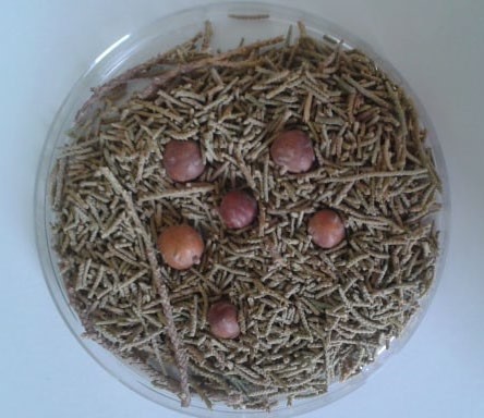
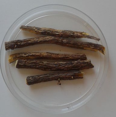
In our study, we used two Algerian medicinal plants, Juniperus phoenicea (Juniper) (Figure 1) and Glycyrrhiza glabra (Licorice) (Figure 2). We have used leaves and fruits (cones) of Juniperus phoenicea which were harvested during February 2019 from the region of Ain Nouissy wilaya of Mostaganem. The roots of Glycyrrhiza glabra have been supplied from the same region. All samples were washed to get rid of debris and all kinds of dust, then they were dried in the open air for fifteen days to obtain a better grinding and keep the substances sought-after bioactive.
|
|
|
Figure 1. Dried leaf of J. phoenicea (used in this work) |
|
|
|
Figure 2. Dried roots of G. glabra (used in this work) |
Extraction and determination of total phenolics
We adopted the protocol described by [7] with slight modifications. 10 g of the powder of the medicinal plants are macerated at room temperature and protected from a light overnight with 100ml of the aqueous solutions: methanol, 70% ethanol (v/v). Filtration was carried out on a piece of muslin fabric, the filtrates were centrifuged at 4000 rpm and room temperature for 20 min. The supernatants were filtered, then concentrated to dryness using a rotavapor. The extracts obtained were stored at 4 °C. The total polyphenol content of the extracts was determined by spectrophotometry according to the colorimetric method by [8].
Results are expressed in mg of gallic acid equivalents per g of dry matter (mg GAE/g dm) according to a gallic acid’s calibration curve.
Total flavonoids assay
Flavonoids were determined according to [9] modified by [10]. 0.5 ml of the sample was mixed with 0.5 ml of 2% AlCl3, after 15 min at room temperature, the absorbance of the sample was measured at 430 nm against the blank prepared from the reagent. Each analysis is repeated three times. Results were expressed as mg of quercetin equivalents per g of dry matter (mg QE/g dm).
Preparation of biological material
The microbial strains used to detect the antimicrobial activity of the prepared extracts of Juniperus phonicea and Glycyrrhiza glabra were original strains from the American Type Culture Collection (ATCC), provided by the LMBAFS laboratory at the University of Mostaganem (Table 1).
Microbial pre-cultures were prepared as described by [11]. Microbial suspensions were prepared 24 hours before inoculation to obtain just confluent colonies. The turbidity was set to 0.5 McFarland that corresponds to 1-2 × 10⁸ CFU/ml for bacteria (OD = 0.08-0.1 at 625 nm), 1-5 × 10⁶ CFU/ml for Candida albicans (OD = 0.12 to 0.15 at 530 nm). Adjusting the inoculum concentration is essential for the quality of the analysis [11, 12].
Antimicrobial activity
The antimicrobial activity test was carried out by two methods: the solid medium diffusion method, in a first step to demonstrate the antimicrobial power of the extracts against the strains used. In the second step, the dilution method in liquid medium (macro-dilution method) was used to measure the minimum inhibitory concentration (MIC) as well as the minimum bactericidal concentration (MBC). The method used is that described by [13, 14].
Table 1. Microbial strains used in this experiment
|
Strain |
Reference |
|
E. coli |
ATCC 25922 |
|
P. aeruginosa |
ATCC 27853 |
|
S. aureus |
ATCC 33862 |
|
C. albicans |
ATCC 10231 |
ATCC: American Type Collection Culture
Solid medium diffusion method
Sterile discs were soaked with 10 μl of each tested extract and placed on the surface of the inoculated medium, then incubated at 37 °C for 24 h for bacteria (E. coli, P. aeruginosa, S. aureus), and 30 °C for 24 h for C. albicans. The inhibition diameter was measured in a centimeter. All experiments were repeated in triplicate.
Micro-dilution method
400 μl of each tested extract was dissolved in dimethylsulfoxide (DMSO) then placed in a sterile tube containing 4.6 ml of Muller Hinton broth medium (BMH). A cascade dilution was carried out in a BMH medium to obtain a concentration range between 80 and 0.3 mg/ml [15]. The MIC of the tested extracts on the strain studied is defined as the lowest concentration that inhibits any bacterial growth visible to the naked eye after 18 or 24 h at 37 °C [11].
13 μl of a microbial inoculum, with a density equivalent to 0.5 Mcfarland (108 CFU/ml), were placed in each of the tubes in the range and were then incubated at 37 °C for 24 h. Control of the microbial growth, for which 13 μl of the adjusted inoculum were deposited in BMH medium added to DMSO was also carried out.
After 24 h of incubation, the MIC of the tested extracts is deduced from the first tube of the range devoid of microbial growth [15].
The minimum bactericidal concentration (MBC) was determined as follows: The same range of concentration, carried out by MIC technique, was used to determine the CMB of the extracts. Samples were taken from each of the tubes devoid of microbial pellet and then deposited "in streaks" on MH agar. The seeded dishes were incubated at 37 ° C for 24 h. The CMB of the extracts was deduced from the streak devoid of bacteria [15].
RESULTS AND DISCUSSION
Variability of molecules extracted from medicinal plants is due to several factors; environmental, geographic and maturation stages of the plant or its components.
Result of the determination of total polyphenols
Total polyphenols amounts of the samples are given in mg GAE/g dm and are reported in Table 2. It was determined that phenolic concentrations of the hydromethanolic extracts were about 122.88±6.64 and 120.54±3.35 mg GAE/g dm for J. phoenicea and G. glabra, respectively.
Hydroethanolic extracts exhibited relatively low phenolic concentrations, about 119.38±7.85 and 110.46±13.34 mg GAE/g dm for J. phoenicea and G. glabra in the same order (Table 1).
Another work done by [16] on the hydromethanolic extract of Glycyrrhiza glabra and has reported that the concentration of the total polyphenols was 118.75 ± 23.68 mg GAE/g, which is lower than (120.54 ± 3.35 mg GAE/g dm) reported in the present study.
[17] reported a total polyphenol concentration of 201±5.8 mg GAE/g for the hydromethanolic extract of J. phoenicea, which was higher than that of the present work (122.88 ± 6.64 mg GAE/g dm).
Flavonoid content
All flavonoid concentrations were calculated from the quercetin calibration range and the results were expressed in mg QE/g dm. Each test was repeated three times as well. Flavonoids concentrations are presented in Table 2.
In general, the content of flavonoids is lower compared to the content of total polyphenols. In Table 2, the highest flavonoid concentrations were recorded to hydromethanolic and hydroethanolic extract of J. phoenicea (15.48 ± 4.97 mg QE/gms and 6.31 ± 1.30 mg QE/ g dm) respectively. In contrast, a concentration of 14.01 ± 8.54 mg QE/gms was recorded belong to the hydromethanolic extract of G. glabra. The lowest concentration (5.16 ± 3.91 mg QE/g dm) was recorded for the hydroethanolic extract of the same species.
Table 2. Total polyphenol and total flavonoids contents of the various extracts
|
Extracts |
Total polyphenols (mg GAE /g dm) |
Flavonoïds (mg QE/g dm) |
|
EHMG |
122.88 ± 6.64 |
15.48 ± 4.97 |
|
EHMR |
120.54 ± 3.35 |
14.01 ± 8.54 |
|
EHEG |
119.38 ± 7.85 |
6.31 ± 1.30 |
|
EHER |
110.46 ± 13.34 |
5.16 ± 3.91 |
EHMG: Hydromethanolic extract of Juniper (Juniperus phoenicea); EHMR: Hydromethanolic extract of Liquorice (Glycyrrhiza glabra); EHEG: Hydroethanolic extracts of Juniper (Juniperus phoenicea); EHER: Hydroethnolic extract of Liquorice (Glycyrrhiza glabra); QE: Quercetin Equivalent; GAE : Gallic acid Equivalent ; dm: dry matter; ±: standard deviation.
It is clearly shown that the highest content of flavonoids belongs to the species Juniperus phoenicea. This difference in the content of flavonoids is probably related to their structural diversity [18] where they are found both in free form or as glycosides [19, 20]. This is depending on the plant species.
In this study, Glycyrrhiza glabra recorded a concentration of 14.01 ± 8.54 mg QE/g dm of the hydromethanolic extract which is a higher value than (0.012 mg QE/g) reported by [21]. The total flavonoid contents of the methanolic and ethanolic extract of Glycyrrhiza glabra showed a large difference in the values with (14.01 ± 8.54 mg QE/g and 5.16 ± 3.91 mg QE/g) respectively.
Result of antimicrobial activity
The present study aims to show the presence or absence of antimicrobial activity, against some pathogenic microbial strains, in the presence of phenolic extracts of Juniperus phoenicea and Glycyrrhiza glabra alone and together.
Result of Solid medium diffusion method
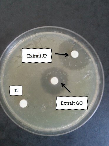
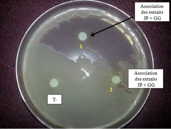
The antimicrobial activity was manifested by the appearance of light areas called zones of inhibition around the discs impregnated with the tested extract (Figure 3).
|
|
|
a) |
|
|
|
b) |
|
Figure 3. Antibacterial effect of JP and GG extracts against Staphylococcus aureus (a: extracts alone; b: combined extract). JP: Juniperus phoenicea extract; GG: Glycyrrhiza glabra extract; T-: Control solution |
|
|
|
Figure 4. Diameters of inhibition areas obtained by hydromethanolic extracts of Juniperus phoenicea and Glycyrrhiza glabra alone and in combination against Staphylococcus aureus. EHMG: Hydromethanolic extract of Juniper (Juniperus phoenicea); EHMR: Hydromethanolic extract of Liquorice (Glycyrrhiza glabra); EC: Combined extract of Juniper and Liquorice (v/v). |
It was observed that the bacterial strain Staphylococcus. aureus was very sensitive to the hydromethanolic extracts of Juniperus phoenicea and Glycyrrhiza glabra with diameters evaluated at (3 ± 0.74 cm and 2.5 ± 0.39 cm) respectively. Therefore, the combined extract of the two medicinal plants gave a much greater effect (diameter 5.5 ± 1.52 cm) (Figure 4).
The strains; Escherichia coli, Pseudomonas aeruginosa, and Candida albicans showed resistance to all extracts studied.
The antibacterial effect of active substances from plant origins mainly depends on the type of bacteria, Gram-positive or negative [22, 23].
The tested extracts exhibited variability depending on the strains studied; where we recorded a complete lack of an antimicrobial effect against the strains (E. coli, P. aeruginosa, and C. albicans), while S. aureus was very sensitive.
As for the hydromethanolic extract of Juniperus phoenicea, the results obtained indicated a significative inhibitory effect (p ≤ 0.05) against Staphylococcus aureus with a diameter inhibition area of 3 ± 0.74 cm. These results are consistent with the results of [24, 25] both have studied phenolic extract effect.
Further studies have been carried out about antibacterial activity against Staphylococcus aureus using herbal extracts. The synergistic effects of Daphne genkwa, Verbena Officinalis, Magnolia Officinalis, and Momordica charantia in combination with gentamicin or oxacillin against methicillin-susceptible (ATCC25923) and methicillin-resistant (ATCC43300) S. aureus were evaluated. This study identified the bioactive ingredients of plants that potentially have antibiotic effects [26].
On the other hand, no antibacterial activity of this extract was reported against E. coli and P. aeruginosa. The extraction method and the nature of the solvent can influence the antibacterial activity of the phenolic compounds of J. phoenicea [22].
No antifungal activity of the three extracts was recorded against Candida albicans. It is difficult to compare these results with those in the literature because the use of different extraction methods reduces the reliability of the comparison between the studies.
The structural difference makes Gram (+) bacteria more sensitive to various natural compounds such as plant extracts [27].
Certain properties of the outer membrane of Gram- bacteria are also assets against the action of antibacterial agents [28, 29]. Therefore, the combined extract in this study has shown no inhibition against Escherichia coli and Pseudomonas aeruginosa (Gram-negative). Another study have indicate that the use of α-pinene-/sabinene-rich juniper EO with anti-QS properties along with refrigeration could provide an additional advantage for controlling the spoilage activities of pseudomonads in fish [30].
Macro-dilution method in liquid medium
The macro-dilution method in liquid medium was performed to determine the MIC and CMB of the hydromethanolic extracts of Juniperus phoenicea and Glycyrrhiza glabra alone and in combination against the strain Staphylococcus aureus. values of the CMB/MIC ratio were also studied (Table 4).
According to our results illustrated in the table above, the lowest MIC is that of the combined extract
(2.5 mg/ml), followed by that of Juniperus phoenicea extract (10 mg/ml) and finally the hydromethanolic extract of Glycyrrhiza glabra (20 mg/ml). The lowest CMB (2.5 mg/ml) belongs to combined extract, in contrast, the MIC of hydromethanolic extracts of Juniperus phoenicea and Glycyrrhiza glabra is identical (80 mg/ml).
MIC is the lowest concentration of inhibitory antibiotic for which it no longer has visible microbial germs. In addition, the bactericidal effect is an effect manifested by an acceleration of the death of bacteria in vivo or in vitro [11].
It was reported that when the CMB/MIC ratio is <4, the extract is considered to be 'bactericidal' [22].
In the present study, the CMB/MIC ratio of the three extracts is greater than 4, which means that the extracts studied have a bacteriostatic effect against Staphylococcus aureus.
G. glabra’s pharmacological activities have been largely determined against various parasites and microorganisms, plasmodium falciparum, and viruses. A study results suggesting that a complex mixture of L. paracasei HP7 containing P. frutescens and G. glabra extracts may be an alternative to treating diseases caused by H. pylori infection [5].
Additionally, it shows anti-inflammatory, antifungal, cytotoxic activities, antioxidant, and anticarcinogenic. In recent study, three active compounds of Gg have the potential to be strong inhibitors for Mpro of SARS-CoV2 but glycyrrhizic acid has a high binding affinity and a good properties [31].
Juniperus extract has a strong antioxidant activity and gastrointestinal effect [32, 33]. Juniperus phoenica L. leaves extract has a good protective role against Gamma-irradiation induced Oxidative stress [34, 35].
Table 3. Results of MIC and CMB of tested extracts against Staphylococcus aureus.
|
|
MIC (mg/ml) |
MBC (mg/ml) |
MBC/MIC |
|
EHMG |
10 |
80 |
8 |
|
EHMR |
20 |
80 |
4 |
|
Combined extract |
2.5 |
20 |
8 |
EHMG: Hydromethanolic extract of Juniper (Juniperus phoenicea); EHMR: Hydromethanolic extract of Liquorice (Glycyrrhiza glabra)
CONCLUSION
As a Mediterranean country, Algeria is a huge source of active molecules of plant origin. For this effect, the aim of this study was of making a modest contribution to solve problems of resistance developed by microorganisms to antimicrobial agents (antibiotics and antifungals).
Our objective was to assay some bioactive molecules of two medicinal plants belonging to Glycyrrhiza glabra, Algerian flora, and Juniperus phoenicea, as well as to test the antimicrobial potential of their extracts.
The micro-constituents of plants, of which polyphenols are the main representatives, provide beneficial effects against the development of various pathologies. These molecules are found in plants, from roots to fruit. The hydromethanolic extraction of phenolic compounds from the plant studied has shown the highest yield 56.15% for Juniperus phoenicea and 47.40% for Glycyrrhiza glabra. This is in comparison to hydroethanolic extraction. polyphenols concentrations of the hydromethanol extract of Glycyrrhiza glabra and Juniperus phoenicea gave respective contents of 122.88 ± 6.64 and 120.54 ± 3.35 mg EAG/g dm.
The antimicrobial potential by the method of diffusion on solid medium (aromatogram), showed a great inhibitory effect of the two hydromethanolic extracts of Juniperus phoenicea and Glycyrrhiza glabra against the pathogenic strain Staphylococcus aureus ATCC 33862. No inhibitory effect on the other strains studied (C. albicans ATCC 10231, P. aeruginosa ATCC 27853, and E. coli ATCC 25922).
From these results, a study of all chemical compositions of the extracts is necessary to identify and specify the active ingredients and to understand their mode of action In Vitro and In vivo, with the aim of more significant data to practice in clinical trials. These two medicinal plants are receiving increased attention due to their rich composition and use as antibiotic agents.
Acknowledgments: We express deep thanks to Pr. A. Riazi for providing pathogenic strains from his laboratory (LMBAFS) . Our thanks to the searcher N. Amara for her practical help.
Conflict of interest: None
Financial support: The entire work was supported by the faculty of Science, nature and life of the university of Mostaganem in Algeria.
Ethics statement: None